A low platelet count gets attention quickly.
Sometimes it should. Sometimes the first problem is not the patient. It is the sample.
That is why, when I see an unexpectedly low platelet count, I do not start by building a clinical explanation immediately. My first question is simpler:
Is this platelet count technically believable?
Before thinking about bleeding risk, marrow failure, immune destruction, consumption, or another cause, I want to know whether the analyzer actually counted the platelets correctly.
That sounds basic. But in practice, platelet counts are among the CBC results most likely to be distorted by clumping, specimen problems, large platelets, or analyzer limitations.
The better sequence is:
Confirm the number first. Then explain the pattern.
Start With the Whole CBC
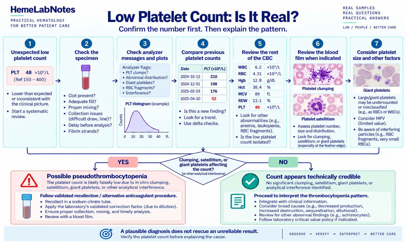
An isolated platelet count should not be read in isolation. If the count is unexpectedly low, I look at the rest of the CBC first. This is the same habit used when reviewing the CBC as a connected pattern.
I want to know:
- Is the hemoglobin stable?
- Is the WBC count unremarkable?
- Are more than one cell line affected?
- Is this a new change?
- Does the analyzer show platelet-related flags?
- Does the specimen look acceptable?
- Does the previous platelet count make sense beside the current result?
A platelet count of 42 × 10⁹/L means something very different when hemoglobin and WBC are stable, yesterday’s platelet count was normal, and the analyzer reports a clump flag than when several cell lines are falling and the platelet count has declined over days.
The number matters. The pattern around the number matters too.
First Question: Could the Specimen Be the Problem?
Platelet interpretation begins before the analyzer result. A poor specimen can create a convincing but false abnormality.
Things I want to know include:
- Is there visible clotting or fibrin?
- Is the tube adequately filled and mixed?
- Was collection difficult?
- Was there a prolonged delay before analysis?
- Does the analyzer report platelet clumping?
- Is the histogram or scatter information unusual?
A small clot can remove platelets from the measurable sample and produce a falsely low count. The analyzer does not know that the specimen no longer represents the patient’s circulating blood. It simply counts what reaches the measurement system.
That is why specimen quality comes before interpretation.
Platelet Clumping: A Common Reason to Stop
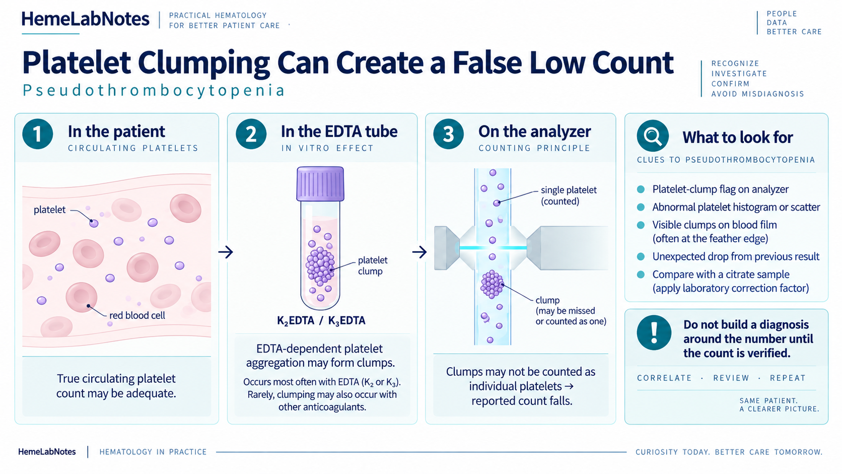
One of the first things I think about with an unexpected low platelet count is platelet clumping.
EDTA-dependent platelet aggregation can produce pseudothrombocytopenia. The patient may have an adequate platelet count in vivo, but platelets aggregate in the collection tube. The analyzer may not recognize those aggregates as individual platelets, and the reported count falls.
This is a laboratory artifact, not true thrombocytopenia. That distinction matters because an unverified false count can lead to unnecessary testing, concern, or intervention.
Analyzer Flags Are Questions, Not Diagnoses
A platelet-clump flag is useful, but it does not diagnose pseudothrombocytopenia. The analyzer is telling us that something about the platelet population does not fit its expected counting pattern.
Depending on the system, platelet-related flags may reflect:
- clumping
- large or giant platelets
- abnormal platelet distribution
- interference in the platelet region
- uncertain separation between platelets and small red-cell fragments
The right response is not simply to copy the flag into the report. It is to ask what the analyzer could not resolve.
Look at the Platelet Histogram or Scatter Information
The numerical count is the final answer generated by the instrument. The histogram or scatter information can show how the analyzer reached that answer.
Clumps, giant platelets, fragments, or interfering particles can distort the expected platelet distribution. Useful warning signs may include an unusual right-hand tail, poor separation from the RBC region, an abnormal scatter distribution, or disagreement between available counting methods.
The exact appearance is platform-specific. The principle is not:
If the number looks wrong, look at the data behind the number.
Compare With Previous Results
Previous platelet counts are one of the fastest ways to decide whether a result deserves closer attention.
Consider an abrupt change:
- Yesterday: 246 × 10⁹/L
- Today: 38 × 10⁹/L
It may be real, but it deserves verification.
Now consider a stable pattern:
- Six months ago: 95 × 10⁹/L
- Three months ago: 91 × 10⁹/L
- Today: 90 × 10⁹/L
The current number is still low, but this is a different laboratory problem. Delta checks should not automatically reject a result. They should make us ask whether the change is biologically and technically plausible.
The Blood Film Should Answer a Specific Question
When clumping is suspected, the peripheral blood film is often the most direct way to resolve the uncertainty. Before looking, I prefer to decide what question I am trying to answer:
Are platelets actually clumped?
or:
Are large platelets present that the analyzer may not have counted reliably?
On the film, I may look for:
- platelet clumps
- platelet satellitism
- large or giant platelets
- a genuine reduction in platelet number
- fragmented red cells
- other morphology that changes the interpretation
The film does not need to answer every clinical question. It should answer the laboratory question created by the CBC.
Platelet Satellitism Can Also Mislead the Count
Platelet satellitism is less common than ordinary clumping, but it is worth recognizing. Platelets may adhere around neutrophils in an EDTA specimen and may not be counted as free platelets by the analyzer.
The automated count can therefore appear lower than the true circulating count. The film is what makes the pattern obvious.
This is another reason not to treat an automated result as infallible when the morphology does not fit.
If Clumping Is Present, Do Not Build a Diagnosis Around the Number
Once platelet clumping is demonstrated, the reported count may no longer represent the patient’s true platelet concentration. The next step should follow the laboratory’s validated procedure.
For suspected EDTA-related clumping, that may include recollection in sodium citrate, repeat analysis, manual review, and an amended or qualified report. If sodium citrate is used, account for dilution using the correction approach validated by the laboratory. A universal correction factor should not be assumed.
Clumping can rarely occur with anticoagulants other than EDTA as well. The recollection and reporting workflow therefore belongs to the local laboratory.
Large Platelets Create a Different Problem
Not every falsely low platelet count is caused by clumping. Large and giant platelets can also affect automated counting.
Very large platelets may fall outside the expected platelet region or be classified differently depending on analyzer methodology. The instrument may exclude them, assign them to another cellular region, or otherwise underestimate the complete platelet population.
This possibility deserves attention when:
- MPV is unavailable or unreliable
- the platelet histogram or scatter is abnormal
- the analyzer flags giant platelets
- the blood film shows large platelets
- the numerical count does not fit the morphology
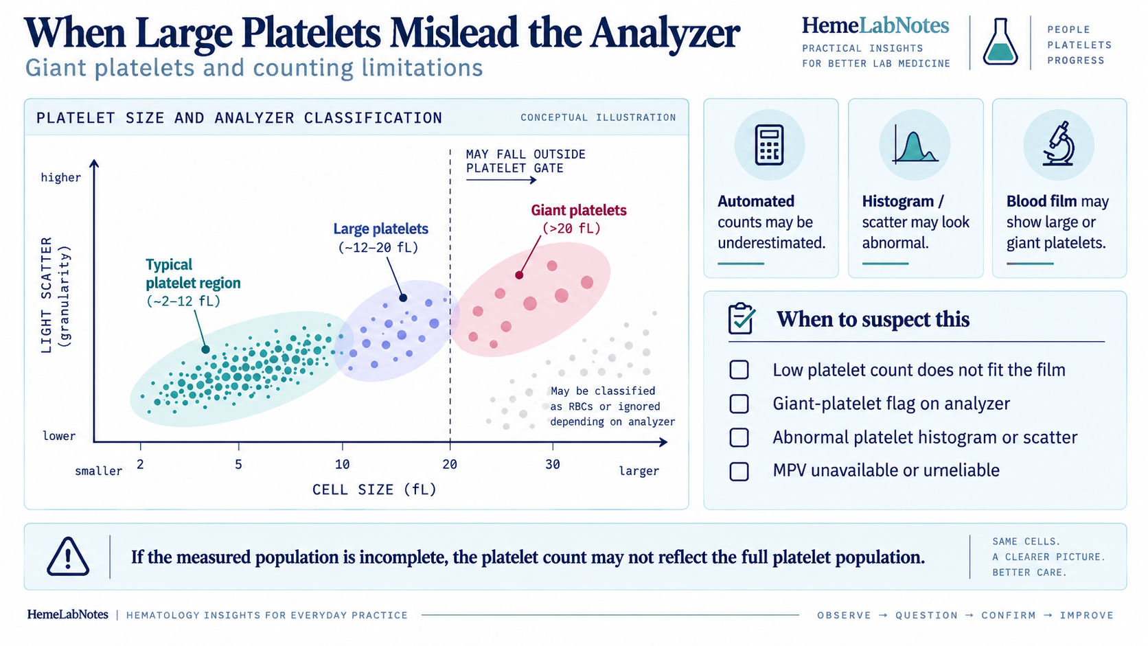
The size boundaries shown in the figure are a conceptual teaching aid, not universal analyzer specifications. Different instruments use different counting principles and discrimination strategies.
MPV Is Not Always Helpful
Mean platelet volume can be useful, but it should not be overinterpreted.
MPV is affected by analyzer methodology, specimen age, anticoagulant, in vitro platelet swelling, and the system’s ability to identify very large platelets. In samples with severe thrombocytopenia, clumping, or giant platelets, MPV may not be reported reliably at all.
If the measured platelet population is questionable, I do not let MPV rescue the interpretation. First establish that the population was measured adequately. Then decide whether MPV adds anything useful.
Platelet Counts Can Be Affected by More Than Platelets
Automated systems separate particles using properties such as size, impedance, light scatter, or fluorescence. Biological particles do not always stay neatly inside the expected categories.
Possible interferences include:
- red-cell fragments
- very small red cells
- cytoplasmic fragments
- platelet clumps
- giant platelets
- other unusual particles
Some interferences may falsely increase the platelet count. Others may decrease it. A number that conflicts with the rest of the CBC deserves investigation.
Is the Low Platelet Count Isolated?
Once I believe the count is technically credible, the question changes:
Is the thrombocytopenia isolated?
An isolated low platelet count and a low count accompanied by anemia and leukopenia are not the same laboratory pattern. This is where the CBC becomes a connected story again.
The clinical explanation comes after technical verification. Broad possibilities may include decreased production, increased destruction, sequestration, consumption, dilution, infection, medication effects, and other clinical processes. The CBC does not settle among them by itself.
Look at the Speed of Change
A platelet count stable around 90 × 10⁹/L for months is different from a sequence of 240, 180, 95, and 38 × 10⁹/L over a short period.
I want to know:
- Is this new?
- Is it worsening or recovering?
- Is there a clear previous baseline?
- Does the clinical timing make sense?
Trend often tells more than one result.
Do Not Forget the Rest of the Film
If thrombocytopenia is genuine, the film can provide information beyond platelet number. Thrombocytopenia with fragmented red cells creates a very different laboratory picture from isolated thrombocytopenia with otherwise unremarkable morphology.
Abnormal white cells, blasts, dysplastic changes, or other significant findings may completely change the direction of investigation. Film review should not stop after finding “platelets decreased.”
The better question is:
What else is happening in this blood sample?
Technical Verification and Urgency May Run in Parallel
Pattern recognition is useful, but severity still matters. A very low platelet count may require prompt action under the laboratory’s critical-value policy.
The practical workflow may therefore have two processes running together:
- Is the result technically credible?
- Is the result clinically urgent according to local policy?
Technical verification should not create unnecessary delay when a result may be urgent. At the same time, urgency does not make an unreliable number accurate.
The threshold and escalation process must come from the laboratory’s validated procedures, not from a universal internet value.
A Bench Example: The Count Is Probably Not Real
Consider:
- Hemoglobin: 13.6 g/dL
- WBC: 7.2 × 10⁹/L
- Platelets: 38 × 10⁹/L
- Previous platelets: 241 × 10⁹/L
- Analyzer flag: platelet clumps
Everything else is relatively stable, and the platelet count has fallen dramatically.
My first thought is not, “What severe disease suddenly caused this thrombocytopenia?” It is:
Is the sample giving me a false platelet count?
If the film shows multiple clumps, I would not build a clinical interpretation around the automated number. That uncertainty needs to be resolved according to laboratory procedure.
Another Bench Example: The Count Looks More Credible
Now consider:
- Hemoglobin: 9.8 g/dL
- WBC: 3.1 × 10⁹/L
- Platelets: 52 × 10⁹/L
- Previous platelets: 68 × 10⁹/L
- No platelet-clump flag or visible clot
- Film shows genuinely reduced platelets
This low count is supported by the previous result, other cytopenias, morphology, and absence of obvious analytical interference.
The problem is no longer mainly, “Is the platelet count real?” It becomes:
What does this broader cytopenic pattern mean?
What If the Count Does Not Match the Film?
This is where I slow down.
If the analyzer reports 25 × 10⁹/L but the film appears to contain more platelets than expected, possible explanations include large platelets, clumping at the feather edge, unusual distribution, analytical interference, or a specimen problem.
Conversely, if the analyzer gives a relatively acceptable count but the film contains obvious clumps, that count may also be unreliable.
The goal is not to decide which result “wins.” The goal is to understand why they disagree.
Platelet clumps may be most obvious toward the feather edge or lateral margins. A quick look at one central field can miss them, so film examination should follow a consistent review pattern.
A Practical Bench Sequence
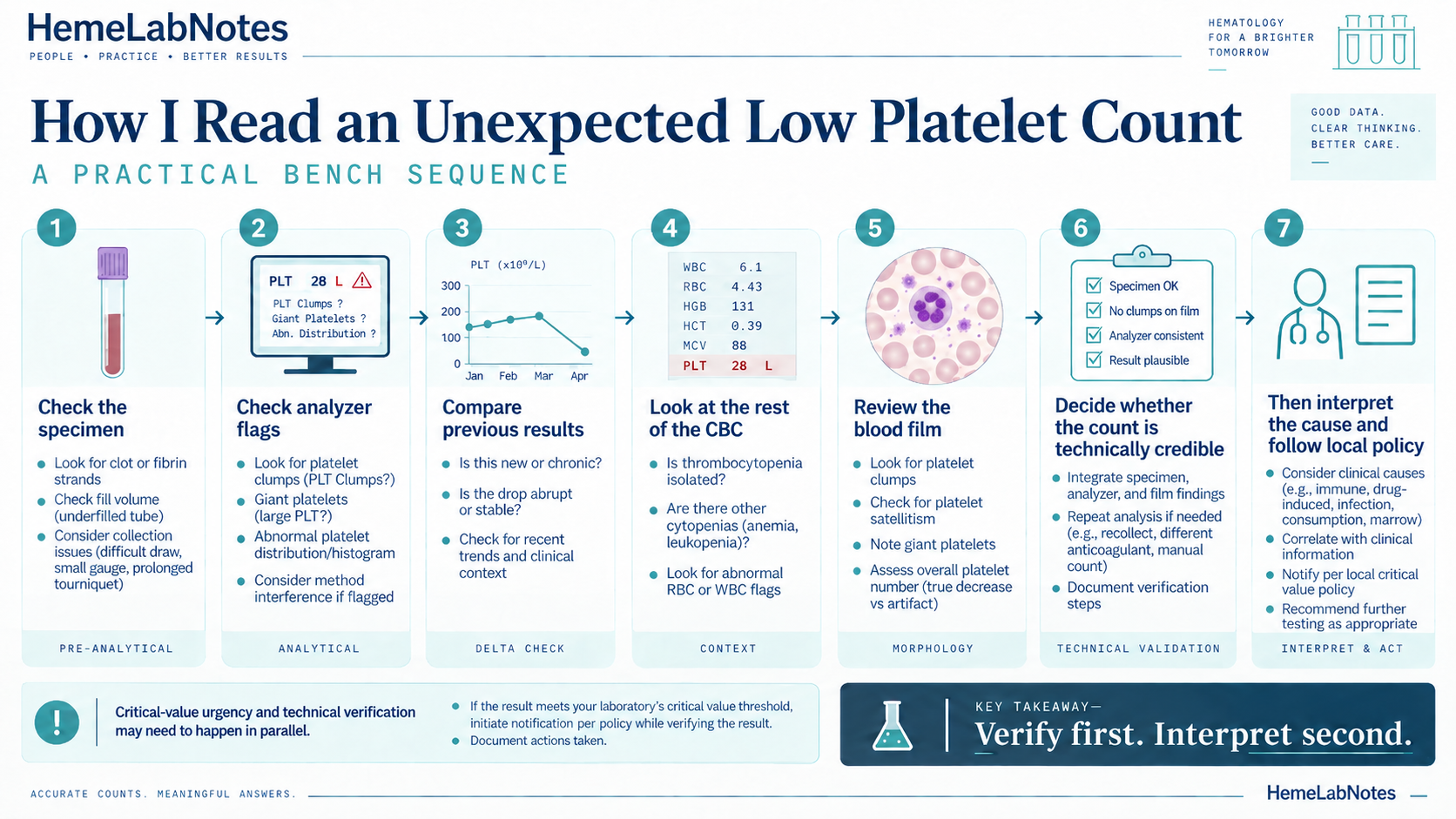
When I see an unexpected low platelet count, I use a sequence like this:
- Check the specimen. Look for clot, fibrin, fill or mixing problems, collection difficulty, delay, and other preanalytical concerns.
- Check analyzer messages and graphics. Review clump flags, distribution warnings, giant-platelet flags, histograms, and scatter information.
- Compare previous results. Ask whether the size and direction of the change make biological and technical sense.
- Read the rest of the CBC. Decide whether the result is isolated or part of a wider cytopenic pattern.
- Review the film when indicated. Look specifically for clumps, satellitism, giant platelets, true reduction, fragments, and significant findings in other cell lines.
- Decide whether the number is technically credible. Integrate the specimen, analyzer, trend, and morphology.
- Interpret and act under validated policy. This may include recollection, citrate testing, repeat or alternative counting, qualified reporting, critical-value notification, and clinical correlation.
The Habit That Improves Platelet Review
When a platelet count looks unexpectedly low, do not immediately explain it. Verify it.
A plausible diagnosis does not rescue an unreliable result. A dramatic number does not become true because it looks clinically important.
The analyzer is very good at counting. Our job is to decide whether it counted the right thing.
Final Thoughts
A low platelet count can be clinically important. It can also be an artifact.
Platelet clumping, specimen clotting, giant platelets, satellitism, and analytical limitations can all distort the automated result. That is why the first question should be:
Is the platelet count real?
Check the specimen. Review the flags and graphics. Look at previous results. Use the blood film when it answers a specific question. Then decide whether the count is technically credible.
Only after that should we start explaining the cause.
Confirm the number first. Then explain the pattern.
Educational Note
This article is intended for laboratory education and professional discussion. Platelet counting methodology, smear-review criteria, citrate correction procedures, alternative-anticoagulant workflows, and critical-value thresholds vary between analyzers and laboratories. Local validated laboratory procedures and clinical policies take precedence.
References
- Cattaneo M. Pseudothrombocytopenia and other conditions associated with spuriously low platelet counts. Haematologica. 2025;110(8):1677–1692. doi:10.3324/haematol.2024.286234.
- Lardinois B, Favresse J, Chatelain B, et al. Pseudothrombocytopenia—A Review on Causes, Occurrence and Clinical Implications. J Clin Med. 2021;10(4):594.
- Bobba RK, Doll DC. Platelet satellitism as a cause of spurious thrombocytopenia. Blood. 2012;119(18):4100.
- Briggs C. Continuing developments with the automated platelet count. Int J Lab Hematol. 2007;29(2):77–91.
- Palmer L, Briggs C, McFadden S, et al. ICSH recommendations for the standardization of nomenclature and grading of peripheral blood cell morphological features. Int J Lab Hematol. 2015;37(3):287–303.
- Bain BJ, Bates I, Laffan MA. Dacie and Lewis Practical Haematology. 12th ed. Elsevier; 2017.
- Bain BJ, Leach M. Blood Cells: A Practical Guide. 7th ed. Wiley; 2025.
- Keohane EM, Preston MM, Mirza KM, Walenga JM, eds. Rodak’s Hematology: Clinical Principles and Applications. 7th ed. Elsevier; 2024.