Sometimes the CBC keeps pointing in one direction, but the next test seems to say very little.
The MCV is low.
The MCH is low.
The RBC count is relatively well preserved.
Iron deficiency does not explain the pattern.
Previous CBCs show the same thing year after year.
Then the hemoglobin analysis comes back looking almost ordinary.
That is where α-thalassemia trait becomes important.
Because unlike classical β-thalassemia trait, there may be no clearly increased HbA₂ to make the pattern obvious.
The absence of an abnormal routine hemoglobin pattern does not erase what the CBC is showing.
A normal-looking hemoglobin analysis does not erase a thalassemia-like CBC pattern.
Why Alpha-Thalassemia Trait Is Easy to Miss
β-thalassemia trait often gives the laboratory another useful clue: increased HbA₂.
α-thalassemia trait is different.
An adult with α-thalassemia trait may have obvious microcytosis and hypochromia while the major hemoglobin fractions remain close to ordinary adult proportions. GeneReviews lists typical adult α-thalassemia trait values of HbA 96–98%, HbF <1%, and HbA₂ 1.5–3.0%. [1]
Those values overlap substantially with ordinary adult hemoglobin fractions.
So the hemoglobin analysis may not look dramatic.
The CBC may be doing most of the talking.
That is why I do not use this shortcut:
Hemoglobin analysis normal → thalassemia excluded
Instead, I ask:
Does a normal-looking hemoglobin analysis actually explain the persistent microcytosis?
If the answer is no, the discrepancy deserves attention.
Start With the CBC Pattern
For HemeLabNotes examples, I continue to use:
- Microcytic: MCV <78 fL
- Normocytic: MCV ≥78 to <98 fL
- Macrocytic: MCV ≥98 fL
These are HemeLabNotes working categories.
Always use your laboratory’s validated reference intervals.
In α-thalassemia trait, the pattern I am looking for is not simply:
low MCV
It is the combination.
For example:
- persistent low MCV
- low MCH
- hemoglobin normal or only mildly reduced
- RBC count preserved or relatively increased
- RDW not dramatically increased unless another process is present
- iron studies that do not adequately explain the microcytosis
- similar previous CBCs
No single feature establishes α-thalassemia trait.
But when several of them travel together, the pattern becomes harder to dismiss.
The RBC Count Still Matters
One reason α-thalassemia trait can look different from uncomplicated iron deficiency is the RBC count.
In iron deficiency, production eventually becomes limited by unavailable iron.
The RBC count may fall.
In α-thalassemia trait, the cells are small because globin-chain production is reduced, but the marrow may maintain a relatively large number of circulating red cells.
GeneReviews notes that RBC count is usually increased in α-thalassemia trait compared with the tendency toward a decreased count in iron deficiency anemia. [1]
That does not mean:
high RBC count = α-thalassemia
It means:
A preserved or relatively high RBC count makes a longstanding thalassemia-type microcytosis more plausible.
Just as in the earlier microcytosis articles, the RBC count changes the question.
It does not answer it.
MCH Is Often Just as Helpful as MCV
MCV gets most of the attention because it tells us that the cells are small.
But MCH is often part of the same pattern.
α-thalassemia trait typically produces both:
- microcytosis
- hypochromia
GeneReviews data illustrate this clearly: α-thalassemia trait has substantially reduced average MCV and MCH compared with unaffected adults. [1]
So when I see a patient with repeatedly low MCV and low MCH, I do not treat them as two independent abnormalities.
I ask what process could produce both, remain stable over time, and still leave the RBC count relatively well preserved.
That combination is often more informative than the MCV alone.
Previous Results May Be More Useful Than Another Formula
A single CBC tells me what is happening today.
Several years of CBCs tell me whether I am looking at a temporary process or a phenotype.
Suppose the current result is:
- MCV 67 fL
- MCH low
- RBC count 5.7 ×10¹²/L
That is interesting.
But if the patient’s CBCs from two, five, and eight years ago show essentially the same pattern, the interpretation changes.
A stable microcytic phenotype makes newly developed isolated iron deficiency less satisfying as the only explanation.
It does not prove α-thalassemia.
But it changes the probability.
This is why I would rather have three old CBCs than three new discrimination indices.
Trend turns a low MCV from a number into a pattern.
RDW Helps — But Do Not Turn It Into a Rule
A relatively stable RDW can support a thalassemia-type pattern.
Iron deficiency often produces more anisocytosis as the deficiency evolves, so RDW may rise more noticeably.
But the overlap is too large to use RDW as a diagnostic divider.
A rapid evidence review similarly notes that normal RDW with microcytosis may suggest thalassemia, but RDW alone lacks sufficient sensitivity or specificity to distinguish thalassemia from iron deficiency. [2]
So I use RDW as another piece of pattern information.
Not as:
normal RDW = thalassemia
or:
high RDW = iron deficiency
Especially because iron deficiency and α-thalassemia can coexist.
Iron Studies Still Have to Fit the Story
Before calling persistent microcytosis a thalassemia-type pattern, iron deficiency has to be addressed properly.
That means more than asking whether somebody once had a normal serum iron.
I want the iron studies to make sense in context.
Depending on the situation, that may include:
- ferritin
- transferrin saturation
- serum iron and transferrin/TIBC where appropriate
- inflammatory context
- previous iron results
- response to treatment if iron deficiency has genuinely been demonstrated
If the iron studies clearly show deficiency, that matters.
But it does not automatically end the investigation.
Iron deficiency and α-thalassemia can coexist. [1]
So sometimes the right interpretation is not:
iron deficiency or α-thalassemia
It is:
iron deficiency on top of an underlying α-thalassemia trait.
That distinction becomes especially useful when iron deficiency is corrected but the MCV never returns to the expected range.
What the Blood Film Adds
The blood film can support the pattern.
Features may include:
- microcytosis
- hypochromia
- anisopoikilocytosis
- target cells
But none of those findings is specific enough to diagnose α-thalassemia trait.
Iron deficiency may also produce microcytosis and hypochromia.
Target cells can occur in several other settings.
And a relatively bland smear does not exclude a carrier state.
So I use the film for the same reason I use the CBC indices:
Does the morphology agree with the rest of the pattern?
The film adds evidence.
It does not provide the genotype. [2]
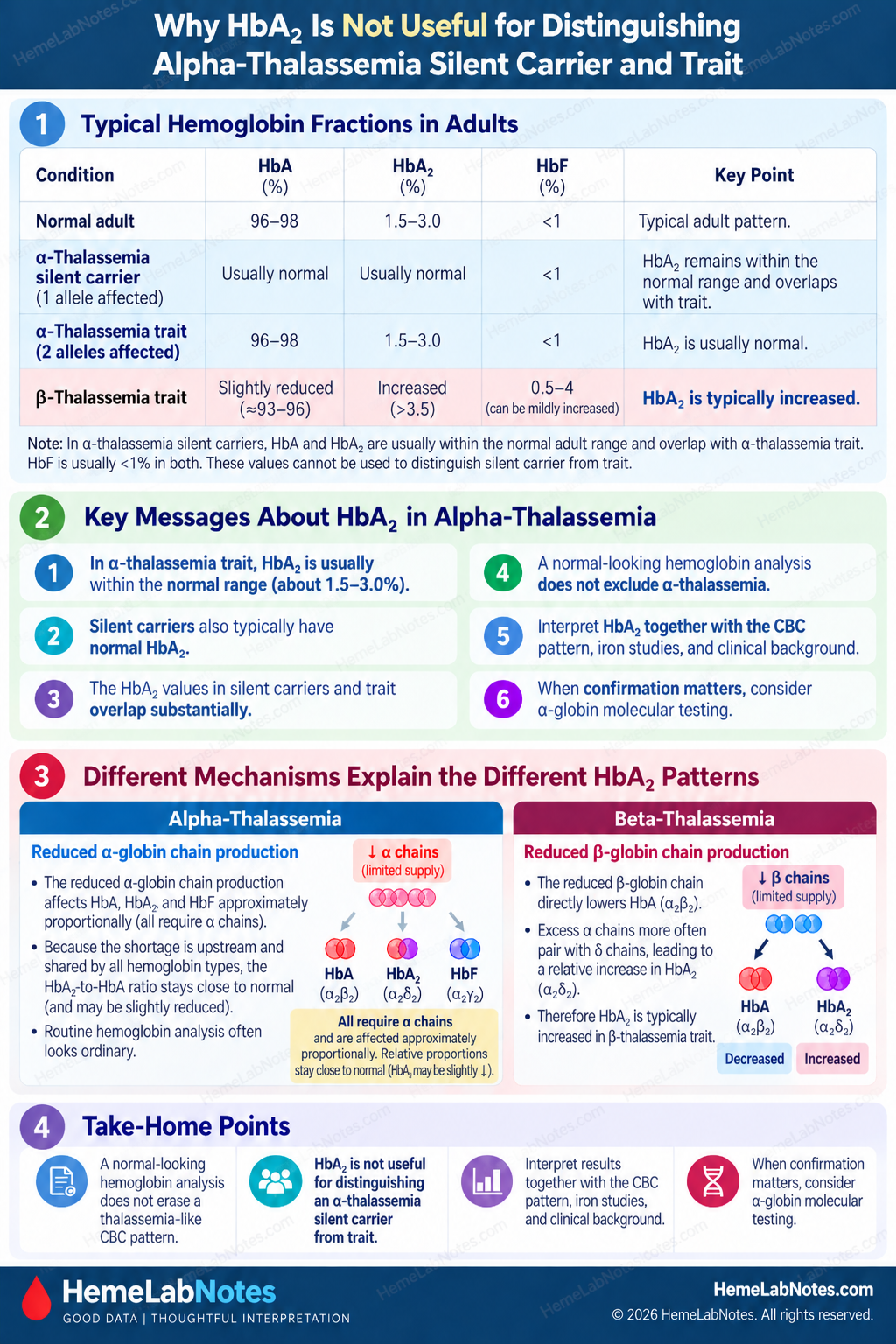
Why HbA₂ May Look Completely Ordinary
This is the part that often causes confusion after a β-thalassemia discussion.
In classical β-thalassemia trait, reduced β-globin production often results in a relative increase in HbA₂.
So HbA₂ becomes a useful screening clue.
α-thalassemia does not work that way.
For α-thalassemia trait, GeneReviews lists typical adult values of:
- HbA: 96–98%
- HbA₂: 1.5–3.0%
- HbF: <1% [1]
Silent carriers may also have an essentially ordinary adult hemoglobin pattern, with HbA₂ remaining within the normal range. [1]
The important point is not a small numerical difference between published carrier-state ranges.
They overlap substantially.
HbA₂ is not a useful way to distinguish an α-thalassemia silent carrier from α-thalassemia trait.
In both states, routine adult hemoglobin analysis may look essentially normal despite an underlying α-globin genotype.
Therefore:
Normal HbA₂ is much less reassuring against α-thalassemia than it is against a classical β-thalassemia-trait pattern.
This is precisely why the CBC and hemoglobin analysis need to be read together.
Silent Carrier and Alpha-Thalassemia Trait Are Not the Same Thing
Humans normally have four functional α-globin alleles.
The phenotype depends largely on how many α-globin alleles are affected and on the type of underlying variant. [1,3]
The simplified spectrum is:
- one affected α-globin allele → α-thalassemia silent carrier
- two affected alleles → α-thalassemia trait
- three affected alleles → HbH disease
- four affected alleles → Hb Bart syndrome
The silent carrier state may have only very mild microcytosis or nearly normal red-cell indices.
Two-allele α-thalassemia trait usually produces a clearer microcytic and hypochromic phenotype. [1,3]
This matters because the number and type of affected α-globin alleles influence the phenotype.
It also explains why the phrase “α-thalassemia carrier” may be too vague when the clinical question requires an exact genotype.
Cis and Trans May Look Similar on the CBC
Two affected α-globin alleles can be arranged in different ways.
They may be on the same chromosome:
–/αα
or one may be affected on each chromosome:
-α/-α
Both represent α-thalassemia trait. [1,3]
From the CBC alone, distinguishing them reliably is not the goal.
But genetically they are not equivalent.
That becomes important when reproductive risk or family testing is the question.
A CBC can tell me that the phenotype deserves investigation.
It cannot tell me with certainty how the affected α-globin alleles are arranged.
The CBC can suggest the phenotype. Molecular testing defines the genotype.
Do Not Expect Routine Hemoglobin Analysis to Find Every Alpha-Thalassemia Carrier
Routine hemoglobin analysis is very useful.
But its limitations matter.
HPLC and capillary electrophoresis measure and separate hemoglobin fractions.
They do not directly count α-globin genes.
A review of molecular α-thalassemia diagnosis emphasizes that CBC, smear, HPLC, and capillary electrophoresis screening cannot detect all α-thalassemia disorders, and molecular analysis is required for confirmatory diagnosis when definitive identification is needed. [4]
That is not a failure of HPLC or capillary electrophoresis.
They are answering a different analytical question.
Hemoglobin analysis measures the hemoglobin phenotype. Molecular testing investigates the globin genotype.
Molecular Testing Is Not One Single Test
It is easy to say:
“Send molecular testing.”
But α-thalassemia itself is genetically heterogeneous.
Many common α-thalassemia variants are deletions.
Others are non-deletional variants.
Different molecular techniques answer different questions.
Depending on the laboratory and population, testing may involve methods designed to identify:
- common α-globin gene deletions
- uncommon or larger deletions
- copy-number changes
- non-deletional sequence variants
Techniques described for α-thalassemia include gap-PCR for common deletions, MLPA for copy-number abnormalities, targeted mutation assays, Sanger sequencing, and increasingly broader sequencing approaches. [4]
The practical point is not that every laboratory needs every method.
It is this:
A negative limited deletion panel is not automatically equivalent to “no α-thalassemia” if the phenotype remains convincing.
What was tested matters.
When Molecular Testing Becomes More Useful
I would think more seriously about α-globin molecular testing when there is:
- persistent unexplained microcytosis
- low MCH that remains unexplained by iron status
- relatively preserved or increased RBC count
- longstanding similar CBC results
- routine hemoglobin analysis that does not explain the CBC
- suspected α-thalassemia where definitive confirmation matters
- family or reproductive implications
- discrepant or complex laboratory findings
Not every mild microcytosis needs immediate genetic testing.
The clinical question matters.
But once the question becomes:
“Does this person carry an α-globin genotype that matters for them or their family?”
CBC pattern recognition is no longer enough.
Three Bench Examples
Example A — Longstanding Microcytosis With Normal Hemoglobin Analysis
Current CBC:
- Hb: 12.1 g/dL
- RBC: 5.7 ×10¹²/L
- MCV: 68 fL
- MCH: 21.2 pg
- RDW: relatively stable
Previous CBCs:
- similar microcytosis over several years
Iron studies:
- ferritin not reduced
- transferrin saturation not reduced
Hemoglobin analysis:
- HbA: approximately 97%
- HbA₂: 2.4%
- HbF: <1%
The hemoglobin analysis is not showing a classical β-thalassemia-trait pattern.
But it also has not explained the microcytosis.
The longstanding low MCV, low MCH, preserved RBC count, and adequate iron status continue to support a thalassemia-type phenotype.
α-thalassemia trait remains a reasonable consideration.
This is the kind of discrepancy where α-globin molecular testing may become useful if confirmation matters.
Example B — Microcytosis That Fits Iron Deficiency Better
CBC:
- Hb: 10.2 g/dL
- RBC: 4.3 ×10¹²/L
- MCV: 71 fL
- MCH: reduced
- RDW: clearly increased
Previous CBC:
- previously normocytic
Iron studies:
- ferritin low
- transferrin saturation low
This is a different pattern.
The microcytosis is new.
The RBC count is not relatively increased.
The RDW has widened.
The iron studies supply a coherent explanation.
α-thalassemia does not have to be invoked simply because the MCV is low.
Microcytosis is a finding. The rest of the pattern tells us what to investigate.
Example C — Alpha-Thalassemia-Type Pattern With New Iron Deficiency
Previous CBC:
- Hb near normal
- RBC count approximately 5.5 ×10¹²/L
- MCV persistently around 69–71 fL
- RDW relatively stable
Current CBC:
- Hb: 10.5 g/dL
- RBC: 5.2 ×10¹²/L
- MCV: 64 fL
- MCH: approximately 20 pg
- RDW: now increased
Current iron studies:
- ferritin low
- transferrin saturation low
This is where historical data matter.
If I looked only at today’s sample, I might concentrate entirely on iron deficiency.
But the older CBCs already showed substantial microcytosis before the current iron deficiency appeared.
Now I have two patterns:
a longstanding microcytic phenotype
plus:
a new iron-deficient change
Treating the iron deficiency is appropriate.
But I would not expect it necessarily to erase the underlying microcytosis.
A Practical Bench Sequence
When I see persistent microcytosis and α-thalassemia becomes a possibility:
1. Confirm the CBC pattern
MCV low?
MCH low?
RBC count preserved or relatively high?
↓
2. Look backward
Is this new or longstanding?
Previous CBCs may be more informative than another index.
↓
3. Review iron status
Does iron deficiency adequately explain the pattern?
Could iron deficiency and thalassemia coexist?
↓
4. Review RDW and the blood film
Do they support the pattern?
Do not use either as a diagnostic rule.
↓
5. Review hemoglobin analysis
Is HbA₂ increased?
Is another variant present?
Does the result actually explain the CBC?
↓
6. If the adult hemoglobin pattern looks ordinary, do not stop automatically
α-thalassemia trait can have HbA, HbA₂, and HbF values close to ordinary adult proportions.
↓
7. Ask what question still needs answering
Is a probable phenotype enough?
Or does the patient need definitive carrier or genotype information?
↓
8. Consider α-globin molecular testing when appropriate
Especially when the phenotype remains convincing or reproductive or family implications make definitive characterization important.
Common Interpretation Traps
Trap 1 — “HbA₂ is normal, so there is no thalassemia”
That logic misses the distinction between β- and α-thalassemia.
Normal HbA₂ does not exclude α-thalassemia trait.
Trap 2 — “The ferritin is low, so everything is iron deficiency”
Iron deficiency and α-thalassemia can coexist.
Previous CBCs can reveal whether the microcytosis existed before the iron deficiency.
Trap 3 — “The RBC count is high, so this must be thalassemia”
A preserved or increased RBC count is a clue.
Not a molecular diagnosis.
Trap 4 — “Normal hemoglobin electrophoresis means normal globin genes”
Hemoglobin analysis examines the hemoglobin phenotype.
It does not directly establish the α-globin genotype.
Trap 5 — “A negative molecular test excludes every alpha-thalassemia variant”
That depends on what the assay actually tested.
A limited common-deletion panel and a comprehensive α-globin investigation are not the same thing.
What the Laboratory Can Say — and What It Cannot
The laboratory can identify a pattern that is compatible with α-thalassemia trait.
It can show persistent microcytosis.
It can show reduced MCH.
It can show a relatively preserved RBC count.
It can demonstrate that iron deficiency does not adequately explain the findings.
It can show that routine adult hemoglobin fractions are unremarkable.
But those findings do not establish the exact α-globin genotype.
That distinction matters.
A careful laboratory interpretation should help the next question become clearer without claiming more than the data support.
Pattern recognition can tell us where to look. Genotype requires the appropriate genetic test.
The Takeaway
α-thalassemia trait is easy to overlook because the CBC may look much more abnormal than the routine hemoglobin analysis.
The pattern becomes more convincing when there is:
- persistent microcytosis
- low MCH
- relatively preserved or increased RBC count
- iron studies that do not explain the finding
- similar previous CBCs
- an adult hemoglobin pattern without the HbA₂ increase expected in classical β-thalassemia trait
A normal-looking HbA₂ does not settle the question.
Neither does a normal-looking HPLC or capillary electrophoresis pattern.
When the CBC keeps pointing toward thalassemia but the routine hemoglobin analysis does not provide the answer, α-thalassemia is one of the important explanations to consider.
And when confirmation matters, the next step is not another CBC formula.
It is asking whether the α-globin genes themselves need to be investigated.
When the CBC keeps pointing toward thalassemia but HbA₂ stays ordinary, think about what routine hemoglobin analysis cannot see.
References
-
Tamary H, Greenberg-Kushnir N, Dgany O. Alpha-Thalassemia. In: Adam MP, Bick S, Mirzaa GM, et al., eds. GeneReviews® [Internet]. Seattle (WA): University of Washington, Seattle; 1993–2026. Updated April 23, 2026.
-
Baird DC, Batten SH, Sparks SK. Alpha- and beta-thalassemia: rapid evidence review. Am Fam Physician. 2022;105(3):272–280.
-
Harteveld CL, Higgs DR. α-thalassaemia. Orphanet J Rare Dis. 2010;5:13. doi:10.1186/1750-1172-5-13.
-
Vijian D, Wan Ab Rahman WS, Ponnuraj KT, Zulkafli Z, Mohd Noor NH. Molecular detection of alpha thalassemia: a review of prevalent techniques. Medeni Med J. 2021;36(3):257–269. doi:10.5222/MMJ.2021.14603.
Further Reading
- Bain BJ, Bates I, Laffan MA. Dacie and Lewis Practical Haematology. 12th ed. Elsevier; 2017.
- Keohane EM, Preston MM, Mirza KM, Walenga JM, eds. Rodak’s Hematology: Clinical Principles and Applications. 7th ed. Elsevier; 2024.