A low MCV may raise the possibility of thalassemia.
A relatively high RBC count may strengthen that suspicion.
Iron studies may make straightforward iron deficiency less convincing.
The blood film may add target cells or other supportive morphology.
But eventually another question appears:
“What does the hemoglobin analysis actually show?”
That sounds simple.
It often is not.
An increased HbA₂ can strongly support β-thalassemia trait.
But a normal HbA₂ does not automatically exclude every thalassemia.
A borderline result can become difficult when iron deficiency, another globin disorder, or an analytical limitation is present.
And sometimes the most useful finding is not a number at all.
It is the fact that the hemoglobin pattern does not match the CBC.
Hemoglobin Analysis Is Not the Starting Point
I would not start with HbA₂ and work backward.
I start with the CBC.
For HemeLabNotes examples, I use:
- Microcytic: MCV <78 fL
- Normocytic: MCV ≥78 to <98 fL
- Macrocytic: MCV ≥98 fL
Always use your own laboratory’s validated reference intervals.
Before looking at the hemoglobin fractions, I want to know:
- How low is the MCV?
- Is MCH also reduced?
- Is the RBC count reduced, preserved, or relatively high?
- Is RDW stable or increasing?
- Is the pattern new or longstanding?
- Do the iron studies explain it?
- What did previous CBCs look like?
Only then do I ask what hemoglobin analysis adds.
The CBC raises the question. Hemoglobin analysis helps refine it.
What HbA₂ Adds
HbA₂ is one of the most useful laboratory clues for β-thalassemia trait.
In a typical β-thalassemia carrier, the pattern includes reduced MCV and MCH with HbA₂ above 3.5%. HbF may also be mildly increased. [1]
So if I have:
- persistent microcytosis
- low MCH
- relatively preserved RBC count
- iron studies that do not explain the microcytosis
- HbA₂ clearly increased
the pattern becomes much stronger.
But there is an important distinction.
HbA₂ supports the pattern. It does not replace the pattern.
A value above a cutoff should not be interpreted without looking at the CBC, iron status, analytical method, and any other abnormal hemoglobin fractions.
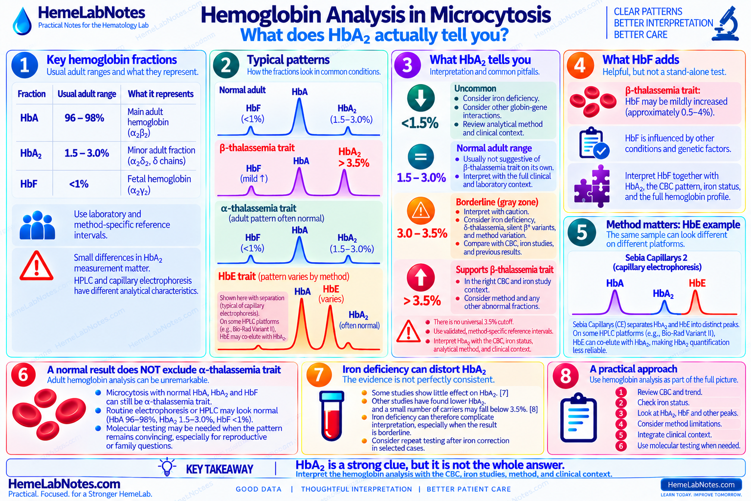
Do Not Treat 3.5% as a Universal Instrument Cutoff
The familiar number is 3.5%.
It is useful.
But it should not become a laboratory law.
GeneReviews uses HbA₂ >3.5% as a typical feature of β-thalassemia carrier status. [1]
At the bench, however, HbA₂ measurement depends on analytical performance and methodology.
ICSH emphasizes that HbA₂ measurement must be precise and accurate because the difference between carrier and non-carrier values is relatively small. HPLC and capillary electrophoresis each have analytical advantages and potential pitfalls. [2,3]
So I prefer:
“Is HbA₂ increased according to this laboratory’s validated method?”
rather than:
“Is it above one universal number?”
Method-specific reference intervals and decision limits matter most when the result sits close to the boundary.
What HbF Adds
HbF sometimes gets less attention because HbA₂ is usually the main screening clue in β-thalassemia trait.
But HbF still adds context.
β-thalassemia carriers may have mildly increased HbF. GeneReviews lists approximately 0.5–4% in carriers. [1]
That can support the pattern.
But I would not diagnose β-thalassemia trait from mildly increased HbF alone.
HbF can be influenced by other conditions and genetic factors, and its interpretation belongs with the rest of the hemoglobin pattern. [4]
So I read HbF as:
Additional information, not a replacement for HbA₂ or the CBC.
A Normal Hemoglobin Analysis Does Not Exclude Alpha-Thalassemia Trait
This is one of the most important practical points.
A patient may have:
- persistent microcytosis
- low MCH
- relatively preserved RBC count
- normal iron studies
- a longstanding pattern
and yet routine adult hemoglobin analysis looks almost unremarkable.
That does not make the CBC irrelevant.
It may make it more important.
For α-thalassemia trait, typical adult values include:
- HbA: 96–98%
- HbF: <1%
- HbA₂: 1.5–3.0% [5]
Those numbers overlap substantially with an ordinary adult pattern.
So:
Normal electrophoresis, HPLC, or capillary electrophoresis does not automatically exclude α-thalassemia trait.
If the CBC continues to show a convincing unexplained microcytic pattern, molecular investigation may still be needed depending on the clinical question. Routine CBC and hemoglobin analysis cannot detect every α-thalassemia genotype. [5,6]
Borderline HbA₂ Is Where Interpretation Gets Harder
A clearly increased HbA₂ in the right CBC pattern is relatively straightforward.
A borderline value is different.
That is where I slow down.
I ask:
- Is there iron deficiency?
- Is the CBC strongly thalassemia-like?
- Is another hemoglobin variant present?
- Could another globin-gene interaction affect HbA₂?
- Is the result close to the analytical decision limit?
- What method generated the result?
- Are previous results available?
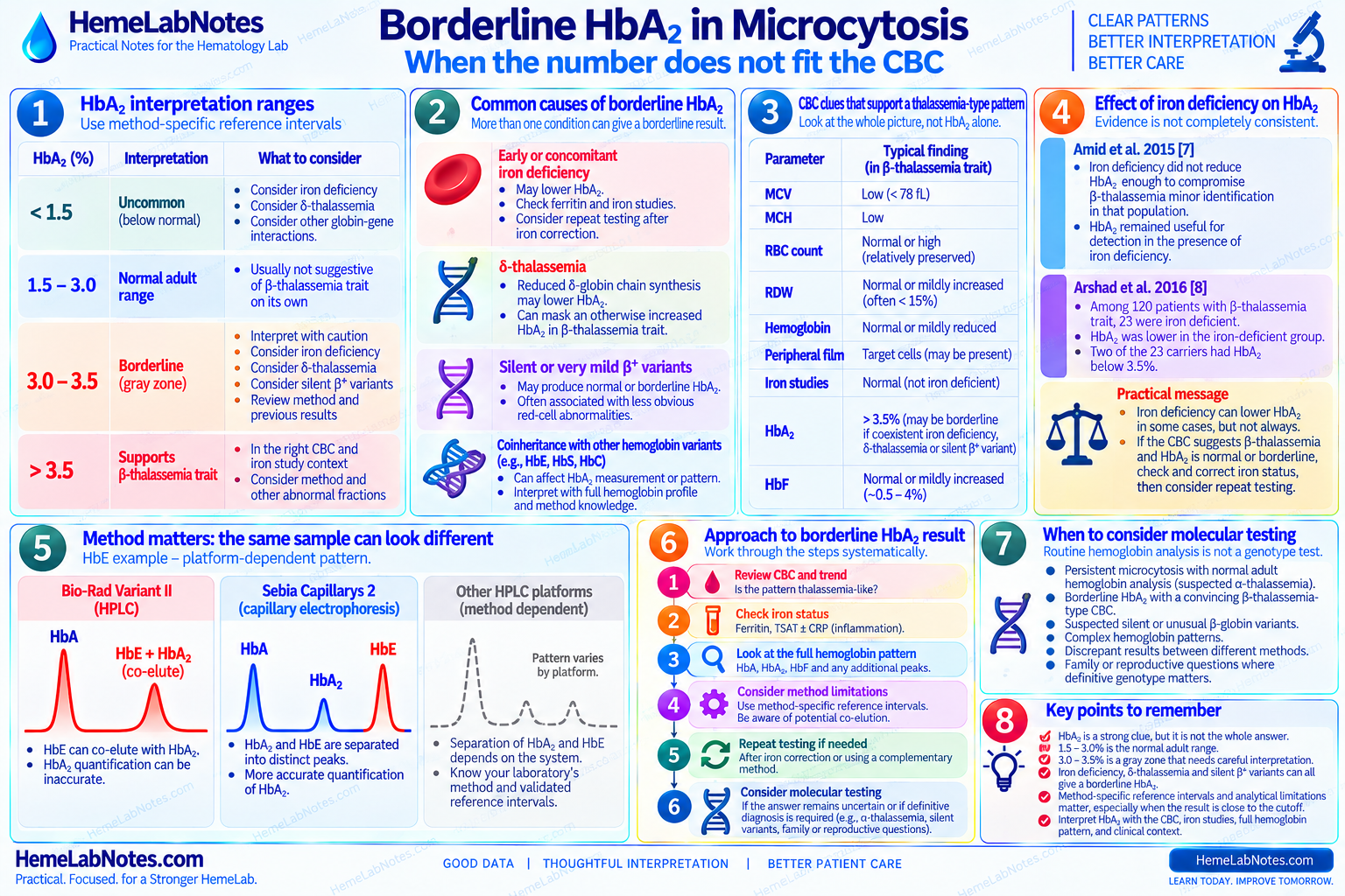
There is no universal borderline interval that applies to every laboratory or method.
Published gray-zone definitions vary.
The important question is whether the value lies close to the laboratory’s validated upper reference or decision limit.
Borderline is not a diagnosis. It is a reason to look harder at the pattern.
Iron Deficiency Can Complicate HbA₂ Interpretation
This is one area where the literature does not give one perfectly tidy answer.
Some studies have found that iron deficiency does not materially prevent detection of β-thalassemia trait using HbA₂.
In one study, iron deficiency did not reduce HbA₂ enough to compromise β-thalassemia minor identification in that population. [7]
But not every study agrees.
In another series of 120 patients with β-thalassemia trait, 23 were iron deficient. HbA₂ was lower in the iron-deficient group, and two of those 23 carriers had HbA₂ below 3.5%. [8]
That is a small number.
But it is enough to make the practical point.
I would not use either of these shortcuts:
iron deficiency present → HbA₂ is unreliable
or:
HbA₂ not increased → β-thalassemia is excluded
Instead:
If the CBC strongly suggests β-thalassemia but HbA₂ is normal or borderline, iron status becomes part of the discrepancy that needs explaining.
Depending on the clinical situation and local laboratory practice, correction of significant iron deficiency followed by repeat hemoglobin analysis may help clarify the pattern.
A Borderline HbA₂ May Have More Than One Explanation
Iron deficiency is not the only possibility.
Coinherited δ-thalassemia can pull an otherwise increased HbA₂ back toward the normal range.
Some silent or very mild β⁺ variants may also produce normal or borderline HbA₂ and less obvious red-cell abnormalities. [1]
This is why I do not read HbA₂ as an isolated yes/no test.
If:
the CBC strongly suggests thalassemia
but:
HbA₂ does not behave as expected
the disagreement itself is useful information.
Do not throw away the CBC because one fraction looks normal.
Look at the Whole Hemoglobin Pattern
Hemoglobin analysis is more than one HbA₂ result.
Depending on the method, the report may contain:
- HbA
- HbA₂
- HbF
- one or more additional peaks or fractions
- retention or migration information
- instrument-specific flags or presumptive windows
The useful question is not simply:
“What is the HbA₂?”
It is:
“Does the entire hemoglobin pattern make sense?”
That includes asking whether an unexpected fraction could be affecting interpretation.
The Chromatogram or Electrophoretic Pattern Matters
This becomes especially important when an abnormal hemoglobin is present.
Automated HPLC and capillary electrophoresis are powerful screening methods.
But neither method resolves every possible variant by itself.
A peak appearing in a particular HPLC window should not automatically be treated as definitive variant identification.
Retention time, peak size, CBC pattern, family or clinical information when relevant, and complementary testing may all matter.
Carrier-screening reviews emphasize that routine hemoglobin methods cannot distinguish every variant completely, and difficult patterns may require DNA confirmation. [9]
So at the bench:
The instrument suggests a pattern. It does not sequence the globin gene.
HbE Is a Good Example of Why the Method Matters
HbE is a useful example because the same specimen can look different depending on the analytical system.
On the Bio-Rad Variant II β-thalassemia program, HbE can co-elute with HbA₂, making HbA₂ difficult to quantify accurately.
Capillary electrophoresis, including Sebia Capillarys systems, can separate HbA₂ and HbE into distinct zones or peaks. [10,11]
That means I would not write a universal rule such as:
“HbE always interferes with HbA₂.”
That is too broad.
The better question is:
“How does our method separate and quantify HbE and HbA₂?”
The HbE issue belongs to the platform, not just to the variant.
Method-Specific Interpretation Is Not a Minor Detail
Two analyzers may both produce a result labeled HbA₂.
That does not mean their analytical behavior is identical.
Comparative work between HPLC and capillary electrophoresis has shown method-dependent differences in HbA₂ quantitation, particularly when structural hemoglobin variants are present. [3,12]
When a result sits close to the laboratory’s decision point, those differences matter more.
That is why a borderline result makes me ask:
- Which platform produced this?
- What is the validated reference interval?
- What is the laboratory’s decision limit?
- Are there known co-elution or migration issues?
- Would a complementary method provide better separation?
- Does the CBC agree?
The closer the number is to the cutoff, the less I want to interpret the number without the method.
Three Bench Examples
Example A — A Classic β-Thalassemia-Type Pattern
- Hb: 12.0 g/dL
- MCV: 66 fL
- MCH: reduced
- RBC count: 5.9 ×10¹²/L
- RDW: relatively stable
- ferritin: not reduced
- TSAT: not reduced
Hemoglobin analysis:
- HbA: predominant
- HbA₂: 5.2%
- HbF: mildly increased
This pattern is internally coherent.
The CBC points toward a thalassemia-type process.
Iron studies do not explain the microcytosis.
HbA₂ strengthens the β-thalassemia carrier interpretation.
The hemoglobin analysis did not create the diagnosis from nothing.
It answered a question that the CBC had already raised.
Example B — Microcytosis but an Unremarkable Adult Hemoglobin Pattern
- Hb: 12.4 g/dL
- MCV: 68 fL
- MCH: reduced
- RBC count: 5.8 ×10¹²/L
- RDW: relatively stable
- iron studies: not iron deficient
- previous CBCs: similar for years
Hemoglobin analysis:
- HbA: 97%
- HbA₂: 2.4%
- HbF: <1%
It would be easy to say:
“Hemoglobin analysis normal — no thalassemia.”
That would go too far.
This pattern does not support classical β-thalassemia trait.
But it does not exclude α-thalassemia trait.
The next question is whether α-globin molecular testing is appropriate for the clinical context.
Example C — Borderline HbA₂ With Iron Deficiency
- Hb: 9.8 g/dL
- MCV: 65 fL
- RBC count: relatively preserved
- RDW: increased
- ferritin: low
- TSAT: low
Hemoglobin analysis:
- HbA₂: near the laboratory decision limit
Now the result is not clean.
The CBC still contains thalassemia-type features.
The iron studies demonstrate iron deficiency.
The HbA₂ is not clearly diagnostic.
I would not force this into:
iron deficiency only
or:
β-thalassemia confirmed
The mixed pattern deserves correlation.
Depending on the circumstances, repeat testing after iron correction or molecular investigation may be appropriate.
When Molecular Testing Becomes Useful
Molecular testing becomes more useful when phenotype and routine screening stop giving a complete answer.
Examples include:
- persistent microcytosis with normal adult hemoglobin analysis and suspected α-thalassemia
- borderline HbA₂ with a convincing β-thalassemia-type CBC
- suspected silent or unusual β-globin variants
- complex hemoglobin patterns
- discrepant HPLC and capillary electrophoresis findings
- family or reproductive questions where definitive genotype matters
Routine hemoglobin methods are excellent screening tools.
They are not universal genotype assays.
For α-thalassemia in particular, molecular analysis is needed when definitive characterization is required because routine CBC, smear, HPLC, or capillary electrophoresis may not detect every genotype. [6]
A Practical Bench Sequence
When I review hemoglobin analysis in a microcytic sample:
1. Start with the CBC
Is the MCV truly low?
How low is the MCH?
What is the RBC count doing?
↓
2. Look at the trend
New microcytosis or longstanding pattern?
↓
3. Review iron status
Does iron deficiency explain the CBC?
Is inflammation complicating ferritin?
↓
4. Look at HbA₂
Clearly increased?
Normal?
Borderline for this method?
↓
5. Look at HbF
Normal or mildly increased?
Does it support the wider pattern?
↓
6. Look at every additional fraction
Unexpected peak?
Variant window?
Unusual migration?
↓
7. Ask what the method can and cannot separate
HPLC and capillary electrophoresis are not analytically identical.
↓
8. Compare everything with the CBC again
Does the hemoglobin pattern explain the red-cell indices?
↓
9. If they disagree, investigate the discrepancy
Do not force the report into the first convenient diagnosis.
↓
10. Consider molecular testing when the answer matters
Especially when α-thalassemia, silent variants, or a complex hemoglobinopathy remains possible.
What Hemoglobin Analysis Does Not Do
Hemoglobin analysis does not make the CBC irrelevant.
It does not exclude α-thalassemia simply because the adult fractions look ordinary.
It does not turn every borderline HbA₂ into β-thalassemia trait.
It does not guarantee that every abnormal peak has been definitively identified.
And it does not remove the need to understand your own analytical method.
That is why I treat it as another layer in the pattern.
Not the whole pattern.
The Takeaway
HbA₂ is one of the strongest routine laboratory clues for β-thalassemia trait.
But the most useful interpretation comes when it agrees with:
- MCV
- MCH
- RBC count
- RDW
- iron studies
- previous CBCs
- HbF
- the rest of the hemoglobin pattern
- the analytical method
A clearly increased HbA₂ can strengthen a β-thalassemia-type pattern.
A normal adult hemoglobin analysis does not automatically exclude α-thalassemia trait.
A borderline HbA₂ should make you look harder at iron status, method limitations, and other globin interactions.
And an unexpected chromatogram should make you ask whether the instrument has shown you the answer — or only the next clue.
Do not ask only whether HbA₂ is “normal.” Ask whether the hemoglobin analysis fits the CBC, iron status, and the rest of the pattern.
References
-
Langer AL. Beta-Thalassemia. In: Adam MP, Bick S, Mirzaa GM, et al., eds. GeneReviews® [Internet]. Seattle (WA): University of Washington, Seattle; 1993–2026. Updated February 12, 2026.
-
Stephens AD, Angastiniotis M, Baysal E, Chan V, Fucharoen S, Giordano PC, Hoyer JD, Mosca A, Wild B; International Council for the Standardisation of Haematology (ICSH). ICSH recommendations for the measurement of haemoglobin A₂. Int J Lab Hematol. 2012;34(1):1–13. doi:10.1111/j.1751-553X.2011.01368.x.
-
Stephens AD, Colah R, Fucharoen S, Hoyer J, Keren D, McFarlane A, Perrett D, Wild BJ; International Council for Standardization in Haematology (ICSH). ICSH recommendations for assessing automated high-performance liquid chromatography and capillary electrophoresis equipment for the quantitation of HbA₂. Int J Lab Hematol. 2015;37(5):577–582. doi:10.1111/ijlh.12413.
-
Stephens AD, Angastiniotis M, Baysal E, Chan V, Davis B, Fucharoen S, Giordano PC, Hoyer JD, Mosca A, Wild B; International Council for the Standardisation of Haematology (ICSH). ICSH recommendations for the measurement of haemoglobin F. Int J Lab Hematol. 2012;34(1):14–20. doi:10.1111/j.1751-553X.2011.01367.x.
-
Tamary H, Greenberg-Kushnir N, Dgany O. Alpha-Thalassemia. In: Adam MP, Bick S, Mirzaa GM, et al., eds. GeneReviews® [Internet]. Seattle (WA): University of Washington, Seattle; 1993–2026. Updated April 23, 2026.
-
Vijian D, Wan Ab Rahman WS, Ponnuraj KT, Zulkafli Z, Mohd Noor NH. Molecular detection of alpha thalassemia: a review of prevalent techniques. Medeni Med J. 2021;36(3):257–269. doi:10.5222/MMJ.2021.14603.
-
Amid A, Haghi-Ashtiani B, Kirby-Allen M, Haghi-Ashtiani MT. Screening for thalassemia carriers in populations with a high rate of iron deficiency: revisiting the applicability of the Mentzer Index and the effect of iron deficiency on HbA₂ levels. Hemoglobin. 2015;39(2):141–143. doi:10.3109/03630269.2015.1024321.
-
Arshad M, Ahmed S, Ali N. Effect of iron deficiency on the phenotype of β-thalassaemia trait. J Coll Physicians Surg Pak. 2016;26(3):230–231.
-
Barrett AN, Saminathan R, Choolani M. Thalassaemia screening and confirmation of carriers in parents. Best Pract Res Clin Obstet Gynaecol. 2017;39:27–40. doi:10.1016/j.bpobgyn.2016.10.015.
-
Munkongdee T, Chen P, Winichagoon P, Fucharoen S, Paiboonsukwong K. Update in laboratory diagnosis of thalassemia. Front Mol Biosci. 2020;7:74. doi:10.3389/fmolb.2020.00074.
-
Chopra P, Bhardwaj S, Negi P, Arora A. Comparison of two high-pressure liquid chromatography instruments Bio-Rad Variant-II and Tosoh HLC-723G11 in the evaluation of hemoglobinopathies. Indian J Hematol Blood Transfus. 2020;36(4):725–732. doi:10.1007/s12288-020-01298-5.
-
Higgins TN, Khajuria A, Mack M. Quantification of HbA₂ in patients with and without β-thalassemia and in the presence of HbS, HbC, HbE, and HbD Punjab hemoglobin variants: comparison of two systems. Am J Clin Pathol. 2009;131(3):357–362. doi:10.1309/AJCP28QKSOPHYOBC.
Further Reading
- Bain BJ, Bates I, Laffan MA. Dacie and Lewis Practical Haematology. 12th ed. Elsevier; 2017.
- Keohane EM, Preston MM, Mirza KM, Walenga JM, eds. Rodak’s Hematology: Clinical Principles and Applications. 7th ed. Elsevier; 2024.