A low MCV often sends the first question toward iron deficiency.
Usually, that is reasonable.
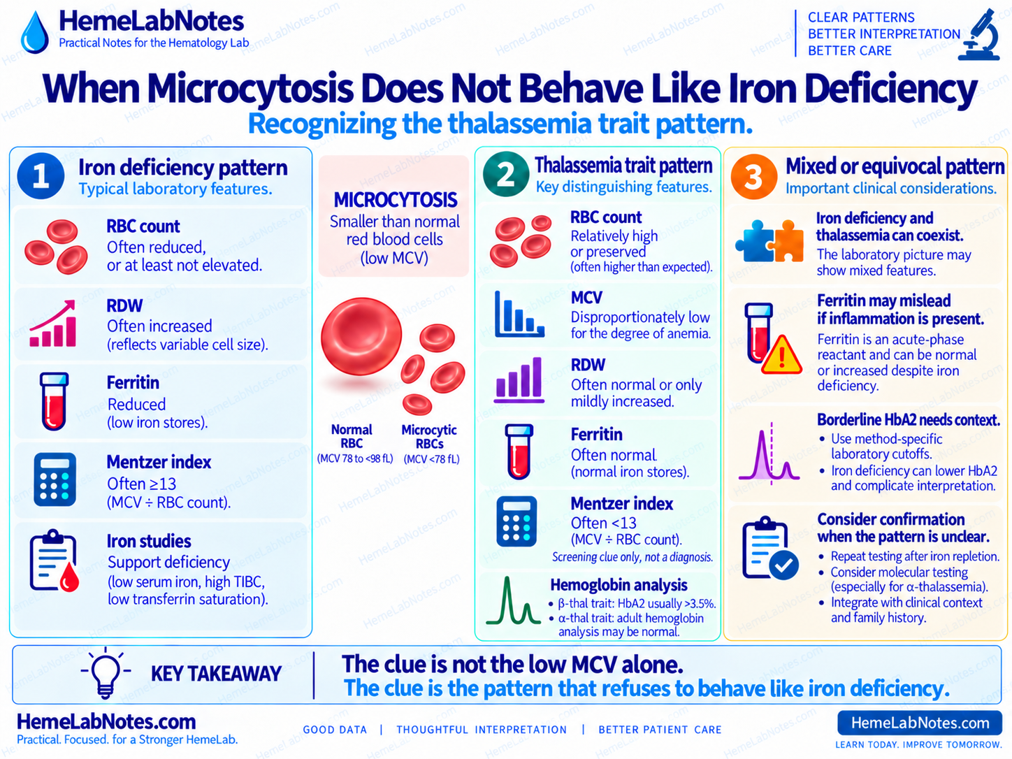
But sometimes the rest of the CBC does not fit.
The MCV may be markedly low.
The hemoglobin may be only mildly reduced.
The RBC count may be preserved or relatively high.
RDW may remain fairly stable.
Iron studies may not support depleted iron stores.
And when previous CBCs are available, the same pattern may have been present for years.
At that point, the useful question changes.
It is no longer:
“Could this be iron deficiency?”
It becomes:
“What else explains this longstanding microcytic pattern?”
That is where a thalassemia-type pattern becomes worth investigating.
Start With the Mismatch
Microcytosis alone is not enough.
For HemeLabNotes examples, I use:
- Microcytic: MCV <78 fL
- Normocytic: MCV ≥78 to <98 fL
- Macrocytic: MCV ≥98 fL
Always use the validated reference intervals from your own laboratory.
Now consider:
- Hb: 12.0 g/dL
- MCV: 67 fL
- RBC count: 5.9 ×10¹²/L
- RDW: relatively stable
The MCV is clearly low.
But the RBC count is not reduced.
The anemia, if present, is mild relative to the degree of microcytosis.
That combination should make you stop and look at the rest of the pattern.
β-thalassemia carriers commonly show microcytosis and reduced MCH, with normal iron studies and increased HbA₂ on quantitative hemoglobin analysis. [1]
Why the RBC Count Matters
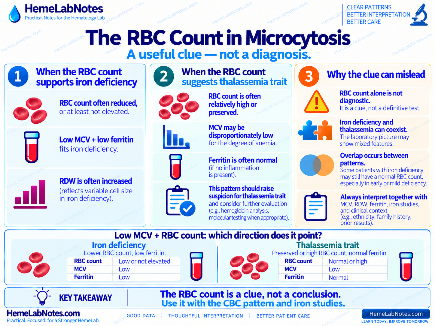
The RBC count is one of the most useful supporting clues in microcytosis.
In straightforward iron deficiency, the RBC count is often reduced, or at least not elevated.
In thalassemia trait, the marrow may continue producing a relatively large number of small red cells.
The result can be:
markedly low MCV + preserved or relatively high RBC count
This is not diagnostic.
But it changes the direction of the workup.
In iron deficiency anemia, the RBC count tends to fall. In α-thalassemia trait, it is usually increased. [2]
So when I see:
- MCV: 68 fL
- RBC count: 5.8 ×10¹²/L
I do not read the RBC count as a separate abnormality.
I ask:
“Does this RBC count fit the rest of the microcytic pattern?”
The Hemoglobin May Be Less Reduced Than Expected
Another clue is the relationship between the degree of microcytosis and the degree of anemia.
A patient may have a very low MCV while hemoglobin remains only mildly reduced or even within the laboratory reference interval.
For example:
Pattern A
- Hb: 12.3 g/dL
- MCV: 66 fL
- RBC count: 5.9 ×10¹²/L
Pattern B
- Hb: 8.5 g/dL
- MCV: 66 fL
- RBC count: 4.1 ×10¹²/L
- RDW: markedly increased
Same MCV.
Different pattern.
For Pattern B:
- calculated Hct ≈ 27.1%
- MCH ≈ 20.7 pg
- MCHC ≈ 31.4 g/dL
That is internally coherent with a microcytic, hypochromic anemia.
Pattern A, by contrast, makes a longstanding thalassemia-type process more relevant.
RDW Helps — But It Is Not a Rule
A relatively stable RDW can support a longstanding, fairly uniform microcytic population.
That often fits better with thalassemia trait than with progressively developing iron deficiency.
But RDW should never become a shortcut.
A normal RDW does not diagnose thalassemia.
A high RDW does not diagnose iron deficiency.
RDW is not sufficiently sensitive or specific to distinguish thalassemia from iron deficiency by itself. [3]
So I use RDW for what it actually tells me:
How uniform is the red-cell population?
The rest of the pattern tells me why that matters.
Previous Results Are Often More Useful Than Another Formula
A single CBC gives you a snapshot.
Several years of CBCs give you behavior.
Three years ago
- Hb: 12.1 g/dL
- MCV: 68 fL
- RBC count: 5.7 ×10¹²/L
- RDW: relatively stable
Last year
- Hb: 12.0 g/dL
- MCV: 67 fL
- RBC count: 5.8 ×10¹²/L
- RDW: relatively stable
Today
- Hb: 12.2 g/dL
- MCV: 68 fL
- RBC count: 5.9 ×10¹²/L
- RDW: relatively stable
That is very different from a patient whose MCV falls from 87 to 80 to 73 to 68 fL while RDW rises and hemoglobin falls.
One pattern is stable.
The other is evolving.
A longstanding abnormality behaves differently from a newly developing one.
This is why previous results often tell me more than another screening calculation.
Iron Studies Should Fit the Story
Before moving toward a thalassemia explanation, ask whether iron deficiency actually explains the microcytosis.
If ferritin is clearly low and the rest of the iron panel supports depleted stores, iron deficiency remains important.
If instead the patient has:
- persistent microcytosis
- relatively high RBC count
- relatively stable RDW
- ferritin not reduced
- TSAT not showing an iron-deficient pattern
- similar CBCs for years
then uncomplicated iron deficiency becomes less convincing.
Thalassemia should be considered when microcytosis is present with normal or elevated ferritin. [3]
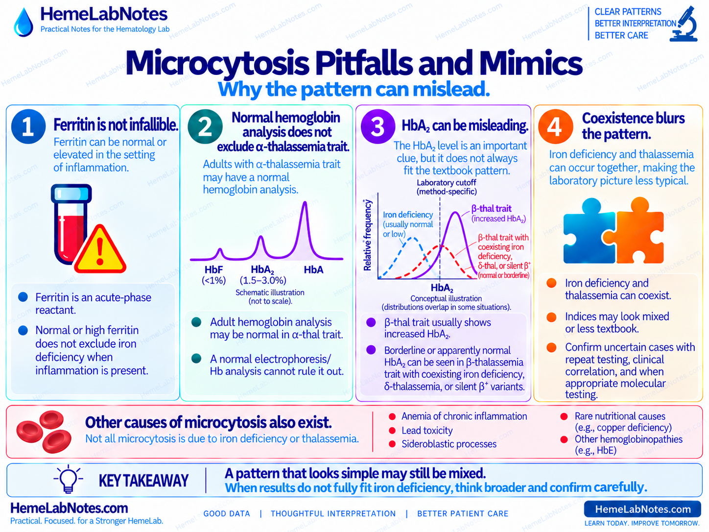
But ferritin has an important limitation.
Ferritin is an acute-phase reactant.
In an inflammatory setting, normal or elevated ferritin does not by itself exclude coexisting iron deficiency. TSAT, transferrin or TIBC, the inflammatory context, and the wider CBC pattern still matter. [4]
So the question is not:
“Is the ferritin normal?”
It is:
“Do the iron studies actually explain the CBC?”
What the Blood Film Adds
The blood film can strengthen the pattern.
Depending on the underlying thalassemia and its severity, findings may include:
- microcytosis
- hypochromia
- target cells
- anisopoikilocytosis
- basophilic stippling
In β-thalassemia, these findings may occur together, but none of them is specific. [3]
The film helps answer:
“Does the morphology agree with the CBC pattern?”
It does not answer:
“Which globin-gene variant does this patient have?”
Morphology remains one layer of evidence.
Mentzer Index: Useful Screening Clue, Not a Verdict
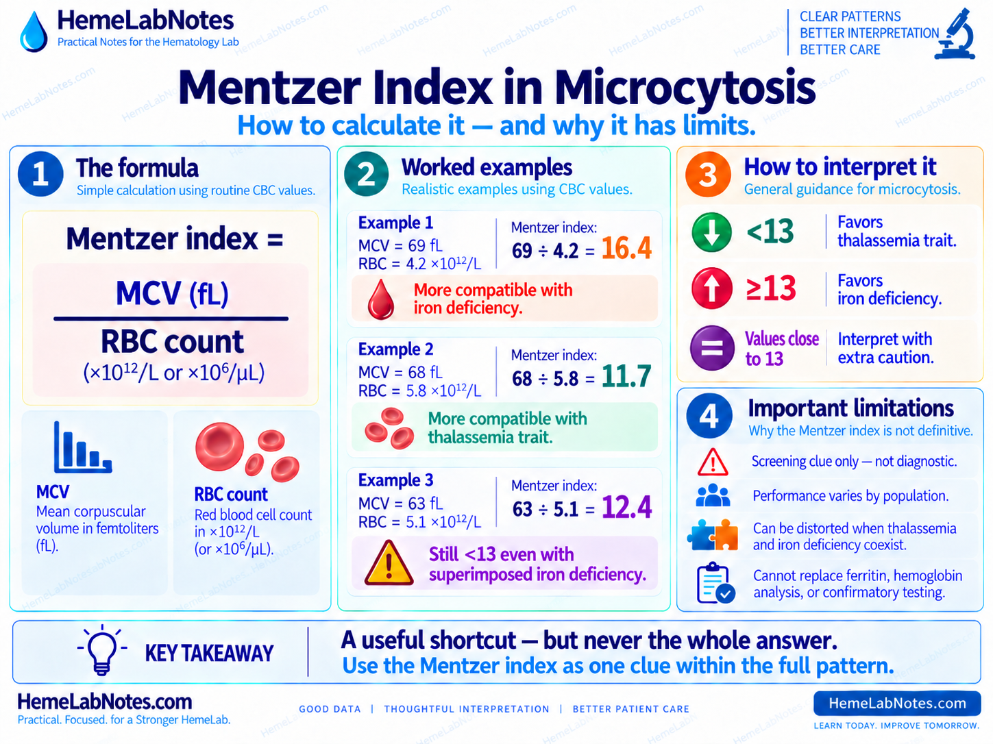
The Mentzer index is:
MCV ÷ RBC count
A commonly quoted screening rule is:
- <13: favors a thalassemia-type pattern
- ≥13: favors iron deficiency
For example:
- MCV: 68 fL
- RBC count: 5.8 ×10¹²/L
68 ÷ 5.8 ≈ 11.7
That supports a thalassemia-type pattern as a screening clue.
But I do not treat 13 as a universal diagnostic cutoff.
The familiar <13 / ≥13 rule is presented for children in the AAFP comparison table, and discrimination indices do not perform equally well across different populations. [3,5]
Adult data make the same point.
In one comparison of six indices, the Green & King index performed best, but none was sensitive enough to safely replace proper investigation. [5]
So I use Mentzer like this:
Supporting direction, not establishing diagnosis.
Hemoglobin Analysis Changes the Question Again
Once iron deficiency no longer explains the pattern well, quantitative hemoglobin analysis becomes important.
For β-thalassemia trait, the typical pattern includes:
- microcytosis
- low MCH
- normal iron studies
- HbA₂ >3.5% [1]
HbF can also be mildly increased. GeneReviews lists approximately 0.5–4% in carriers. [1]
For me, HbA₂ remains the more useful screening clue here.
Laboratories should still use their own validated, method-specific interpretive limits.
So a pattern such as:
- low MCV
- low MCH
- relatively preserved RBC count
- iron studies not showing deficiency
- increased HbA₂
is substantially more informative than any of those findings alone.
But α-thalassemia behaves differently.
A Normal Hemoglobin Analysis Does Not Exclude Alpha-Thalassemia Trait
This is an important bench point.
In adults with α-thalassemia trait, routine hemoglobin analysis can look essentially unremarkable.
Typical values include: [2]
- HbA: 96–98%
- HbF: <1%
- HbA₂: 1.5–3.0%
Those values overlap substantially with the usual adult hemoglobin pattern.
So routine electrophoresis or HPLC may not provide an obvious abnormal fraction pattern.
Normal hemoglobin analysis does not automatically exclude α-thalassemia trait.
If the CBC pattern remains convincing, molecular testing may be appropriate depending on the clinical question, family history, reproductive implications, and local testing strategy. [2,3]
A Normal or Borderline HbA₂ Also Needs Context
HbA₂ is very useful in β-thalassemia carrier detection.
But it is not immune to biological complexity.
Coinherited δ-thalassemia can pull an otherwise increased HbA₂ back toward the normal range.
Some silent β⁺ variants may also present with normal red-cell indices and normal or borderline HbA₂. [1]
Iron deficiency adds another layer of uncertainty.
And the literature does not give us one perfectly tidy answer.
In one study, iron deficiency did not significantly lower HbA₂ in β-thalassemia minor and did not prevent the Mentzer index from separating carriers from individuals without β-thalassemia minor. [6]
But not every study agrees.
In a smaller series, iron deficiency lowered HbA₂ in some carriers. Among 23 iron-deficient patients with β-thalassemia trait, two had HbA₂ below 3.5%. [7]
That is uncommon, but it matters.
A patient can have a convincing β-thalassemia-type CBC pattern and still produce a normal or borderline HbA₂ when another factor is interfering.
So when I see:
- substantial iron deficiency
- a convincing β-thalassemia-type CBC pattern
- normal or borderline HbA₂
I do not automatically close the case.
Depending on local practice and the clinical question, correcting the iron deficiency and reassessing the hemoglobin analysis may be reasonable.
The practical rule is:
Do not interpret a borderline HbA₂ in isolation.
Read it with:
- CBC pattern
- iron status
- analytical method
- previous results
- relevant family history
- laboratory interpretive guidance
Iron Deficiency and Thalassemia Trait Can Coexist
A patient with a longstanding thalassemia-type pattern can still become iron deficient.
That can make a previously neat pattern much less tidy.
Previous baseline
- Hb: 12.1 g/dL
- MCV: 68 fL
- RBC count: 5.8 ×10¹²/L
- RDW: relatively stable
Later
- Hb falls
- MCV falls further
- RDW rises
- ferritin falls
- TSAT falls
- RBC count becomes less preserved
The patient did not stop having a thalassemia trait.
Another process was added.
Iron deficiency and α-thalassemia can coexist, making the laboratory pattern harder to interpret. [2]
A stable inherited pattern can still acquire a new deficiency.
This is another reason previous results matter.
Do Not Treat the CBC as a Genetic Test
One of the easiest shortcuts is:
low MCV + high RBC count = thalassemia
That goes too far.
The same applies to:
- target cells
- stable RDW
- Mentzer index <13
- normal ferritin
Each finding strengthens or weakens a pattern.
None establishes the genotype.
CBC indices are screening clues.
Morphology provides supporting evidence.
Iron studies test an important competing explanation.
Hemoglobin analysis may identify a characteristic pattern.
Molecular testing may be needed when routine hematologic and hemoglobin testing do not provide the answer. [1–3]
Three Bench Examples
Example A — The Pattern Fits Iron Deficiency Better
- Hb: 9.0 g/dL
- MCV: 69 fL
- RBC count: 4.2 ×10¹²/L
- RDW: 18.6%
- ferritin: low
- TSAT: low
- TIBC: increased
Derived values:
- Hct ≈ 29.0%
- MCH ≈ 21.4 pg
- MCHC ≈ 31.0 g/dL
Previous CBCs show progressive decline in MCV and hemoglobin.
Film:
- microcytosis
- hypochromia
- anisocytosis
- pencil cells
The CBC, trend, morphology, and iron studies all point in the same direction.
There is no reason to force thalassemia into the interpretation simply because the MCV is low.
Example B — Longstanding Thalassemia-Type Pattern
- Hb: 12.2 g/dL
- MCV: 67 fL
- RBC count: 5.9 ×10¹²/L
- RDW: relatively stable
- ferritin: not reduced
- TSAT: not reduced
- previous CBCs: similar for years
Mentzer index:
67 ÷ 5.9 ≈ 11.4
Film:
- marked microcytosis
- relatively uniform population
- hypochromia
- target cells
This pattern makes thalassemia trait worth investigating.
But ferritin that is not reduced is reassuring only when the clinical context does not suggest inflammation or another condition that could raise ferritin.
The CBC pattern does not by itself tell us whether the process is α- or β-thalassemia.
Example C — A Longstanding Pattern Has Changed
Previous baseline:
- Hb: 12.1 g/dL
- MCV: 68 fL
- RBC count: 5.8 ×10¹²/L
- RDW: relatively stable
Today:
- Hb: 9.9 g/dL
- MCV: 63 fL
- RBC count: 5.1 ×10¹²/L
- RDW: 18.2%
- ferritin: low
- TSAT: low
The previous CBC suggests a longstanding thalassemia-type baseline.
The current iron studies support superimposed iron deficiency.
Now calculate the Mentzer index:
63 ÷ 5.1 ≈ 12.4
It is still below 13.
If I looked at Mentzer alone, this case would still point toward a thalassemia-type pattern even though the iron studies clearly show that iron deficiency has been added.
That is exactly the problem.
A discrimination index may help separate clean textbook patterns.
It is much less useful when two processes are happening at the same time.
Both processes may be present.
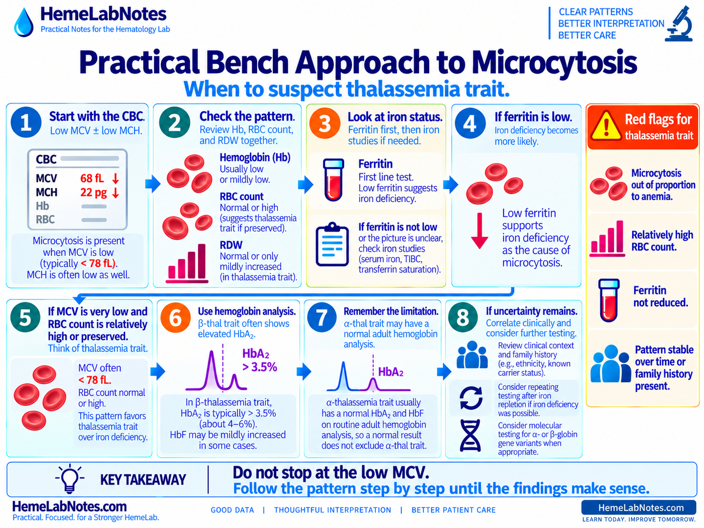
A Practical Bench Sequence
When microcytosis does not fit straightforward iron deficiency:
1. Confirm the MCV
Is the microcytosis real?
↓
2. Look at the hemoglobin
How much anemia is actually present?
↓
3. Look at the RBC count
Reduced, preserved, or relatively high?
↓
4. Add the RDW
Stable population or increasing heterogeneity?
↓
5. Compare previous CBCs
New, progressive, or longstanding?
↓
6. Review iron studies
Do they genuinely support depleted iron stores?
Could inflammation be making ferritin harder to interpret?
↓
7. Review the blood film when indicated
Does morphology support the same pattern?
↓
8. Use discrimination indices cautiously
Mentzer and similar indices can support the direction of investigation.
They do not establish the diagnosis.
↓
9. Review quantitative hemoglobin analysis
Is HbA₂ increased?
Is the result borderline?
Could iron status or another globin disorder affect interpretation?
↓
10. Remember α-thalassemia
A normal adult hemoglobin analysis does not exclude α-thalassemia trait.
↓
11. Consider molecular testing when appropriate
Especially when the laboratory pattern remains convincing but routine hemoglobin testing does not provide the answer.
↓
12. Ask the final question
Not:
“Is the MCV low enough for thalassemia?”
But:
“What explanation best fits the entire pattern?”
The Takeaway
Thalassemia trait is not diagnosed from:
- a low MCV
- a high RBC count
- target cells
- a stable RDW
- a Mentzer index below 13
Those are clues.
What matters is whether several independent pieces point in the same direction:
- marked microcytosis
- relatively preserved RBC count
- compatible RDW
- iron studies that do not explain the microcytosis
- a longstanding pattern
- supportive morphology
- appropriate hemoglobin and/or molecular testing
And one more point matters:
Microcytosis that does not fit iron deficiency is not automatically thalassemia.
Other causes remain possible, including anemia of inflammation, lead-related disorders, sideroblastic processes, other hemoglobinopathies and, less commonly, disorders such as copper deficiency. [3]
The laboratory’s job is not to force the first plausible label onto the CBC.
It is to keep narrowing the question until the pattern makes sense.
The clue is not the low MCV. The clue is the pattern that refuses to behave like iron deficiency.
References
-
Langer AL. Beta-Thalassemia. In: Adam MP, Bick S, Mirzaa GM, et al., eds. GeneReviews® [Internet]. Seattle (WA): University of Washington, Seattle; 1993–2026. Initial posting September 28, 2000. Updated February 12, 2026.
-
Tamary H, Greenberg-Kushnir N, Dgany O. Alpha-Thalassemia. In: Adam MP, Bick S, Mirzaa GM, et al., eds. GeneReviews® [Internet]. Seattle (WA): University of Washington, Seattle; 1993–2026. Initial posting November 1, 2005. Updated April 23, 2026.
-
Baird DC, Batten SH, Sparks SK. Alpha- and beta-thalassemia: rapid evidence review. Am Fam Physician. 2022;105(3):272–280.
-
Fertrin KY. Diagnosis and management of iron deficiency in chronic inflammatory conditions (CIC): is too little iron making your patient sick? Hematology Am Soc Hematol Educ Program. 2020;2020(1):478–486. doi:10.1182/hematology.2020000132.
-
Ntaios G, Chatzinikolaou A, Saouli Z, et al. Discrimination indices as screening tests for beta-thalassemic trait. Ann Hematol. 2007;86(7):487–491. doi:10.1007/s00277-007-0302-x.
-
Amid A, Haghi-Ashtiani B, Kirby-Allen M, Haghi-Ashtiani MT. Screening for thalassemia carriers in populations with a high rate of iron deficiency: revisiting the applicability of the Mentzer Index and the effect of iron deficiency on HbA₂ levels. Hemoglobin. 2015;39(2):141–143. doi:10.3109/03630269.2015.1024321.
-
Arshad M, Ahmed S, Ali N. Effect of iron deficiency on the phenotype of β-thalassaemia trait. J Coll Physicians Surg Pak. 2016;26(3):230–231.
Further Reading
- Bain BJ, Bates I, Laffan MA. Dacie and Lewis Practical Haematology. 12th ed. Elsevier; 2017.
- Keohane EM, Preston MM, Mirza KM, Walenga JM, eds. Rodak’s Hematology: Clinical Principles and Applications. 7th ed. Elsevier; 2024.