A low MCV may point toward iron deficiency.
It does not prove it.
The CBC gives us the pattern.
Iron studies test whether that explanation actually fits.
That sounds straightforward until ferritin is normal, serum iron is low, transferrin saturation is reduced, and inflammation is sitting quietly in the background.
Then the individual numbers stop being enough.
The useful question becomes:
Do the iron studies tell the same story as the CBC?
Start With the CBC Pattern
Before opening the iron panel, look at what brought you there.
For example:
- Hb decreased
- MCV 70 fL
- RDW increased
- RBC count relatively lower
- previous MCV gradually falling
That pattern makes iron restriction a reasonable question.
Now the iron studies have a job:
support it, weaken it, or show that the story is more complicated.
Do not start with the ferritin and work backward toward the CBC.
Start with the pattern.
Then ask whether the biochemical results agree.
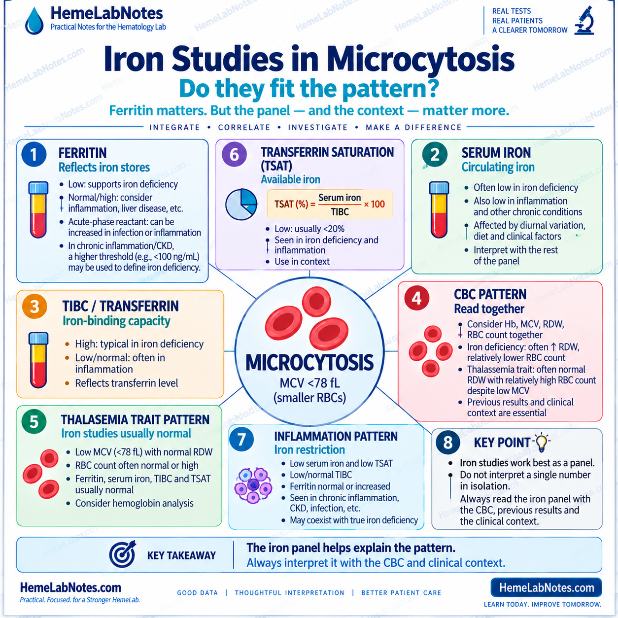
Ferritin Is Usually the First Number I Look At
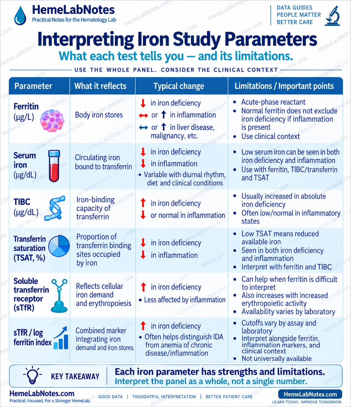
Ferritin is useful because it reflects iron stores.
When ferritin is clearly low, depleted iron stores become much more likely.
That is one of the cleaner patterns we see in laboratory medicine.
The problem begins when ferritin is not low.
Ferritin is also an acute-phase reactant. Infection, inflammation, liver disease, and other clinical settings can increase it, so a normal or elevated ferritin does not automatically exclude iron deficiency. [1,2]
So:
Low ferritin is very useful. Normal ferritin is not always reassuring.
The number still needs context.
Pattern 1: The Iron Studies Fit
Consider:
- Hb: 9.4 g/dL
- MCV: 69 fL
- RDW: 18.4%
- RBC count: 4.4 ×10⁶/µL
- ferritin: low
- serum iron: low
- TIBC: increased
- transferrin saturation: low
This CBC is internally coherent as a microcytic-hypochromic pattern.
The CBC and iron studies are also pointing in the same direction.
In straightforward absolute iron deficiency, ferritin and serum iron typically fall, while transferrin or TIBC tends to rise. The result is reduced transferrin saturation. [2,3,6]
At this point, the question changes.
It is no longer simply:
“Is iron deficiency possible?”
It becomes:
“Why is this patient iron deficient?”
Blood loss?
Reduced intake?
Malabsorption?
Increased requirement?
The CBC identified the pattern.
The iron studies made the explanation more credible.
Now the cause matters.
Serum Iron Is the Number I Trust Least by Itself
Serum iron attracts attention because it looks direct.
Low iron.
Therefore iron deficiency.
Not necessarily.
Serum iron can also fall in inflammation and varies with physiological and clinical conditions.
Both absolute iron deficiency and anemia of inflammation can therefore show low circulating iron. [2,4]
So when serum iron is low, I do not stop there.
I ask:
- What is the ferritin?
- What is the TIBC or transferrin?
- What is the transferrin saturation?
- Is inflammation present?
- Does the CBC pattern support iron deficiency?
Serum iron is a clue inside the panel, not the panel itself.
TIBC and Transferrin Add Direction
TIBC reflects the blood’s iron-binding capacity and is closely related to transferrin.
In straightforward absolute iron deficiency, transferrin production often increases.
That tends to push TIBC upward.
Inflammation can move the pattern in the opposite direction. Transferrin may fall, so TIBC may be lower rather than increased. [2,4]
That distinction matters because serum iron may be low in both situations.
So:
low serum iron + increased TIBC
does not tell the same story as:
low serum iron + low or normal TIBC
Same low serum iron.
Different pattern.
Different next question.
Transferrin Saturation Tells Us About Available Iron
Transferrin saturation — TSAT — tells us how much of transferrin’s iron-binding capacity is actually occupied.
A low TSAT means circulating iron availability is reduced.
Useful.
But still not disease-specific.
TSAT may be reduced in both absolute iron deficiency and inflammatory iron restriction. In inflammatory states, ferritin may remain normal or increased while TSAT falls. [2,4]
So I do not read:
TSAT low = iron deficiency
I read:
TSAT low = available iron is reduced. Why?
Pattern 2: Low Iron, but Ferritin Is Not Low
Consider:
- Hb: 10.4 g/dL
- MCV: 76 fL
- serum iron: low
- transferrin saturation: low
- TIBC: low or normal
- ferritin: normal or increased
- inflammatory markers: increased
This is microcytic by the HemeLabNotes example range of MCV <78 fL.
But it is no longer the clean pattern of uncomplicated absolute iron deficiency.
Inflammation changes iron handling.
Hepcidin rises.
Iron becomes less available to developing red cells.
Iron is retained within storage and reticuloendothelial compartments.
Serum iron falls.
TSAT falls.
Ferritin may remain normal or increase.
TIBC may also be reduced rather than increased. [2,4]
The question is not:
“Is ferritin normal?”
It is:
“Does the ferritin make sense in this clinical setting?”
Normal Ferritin Does Not Always Mean Normal Iron Stores
This deserves its own pause.
Ferritin behaves differently when inflammation is present.
In otherwise uncomplicated settings, ferritin is a strong marker of iron stores.
But when infection or inflammation enters the picture, interpretation becomes less straightforward. WHO specifically recommends considering inflammation when ferritin is used to assess iron status. [1]
That means:
normal ferritin + low TSAT + inflammation
cannot be read in the same way as:
normal ferritin + normal TSAT + no inflammatory context
Same ferritin.
Different story.
Pattern 3: The CBC Looks Iron-Restricted, but the Panel Does Not
Consider:
- Hb: 11.2 g/dL
- MCV: 68 fL
- RDW: relatively stable
- RBC count: 5.7 ×10⁶/µL
- ferritin: not reduced
- transferrin saturation: not clearly reduced
- previous CBCs: almost unchanged for years
At first glance, the low MCV may tempt us toward iron deficiency.
But the rest of the pattern is pushing back.
The RBC count is preserved.
RDW is stable.
The pattern is longstanding.
The iron studies do not support straightforward iron depletion.
That is the point where I stop trying to make iron deficiency fit.
A thalassemia-type process becomes more relevant.
When the biochemical results disagree with the first CBC impression, change the question.
The ARUP microcytic anemia algorithm follows the same general logic: iron assessment comes first, and when iron parameters do not explain the microcytosis, RBC count and hemoglobinopathy evaluation become more relevant. [5]
Iron Deficiency and Inflammation Can Coexist
Real patients do not always choose one mechanism.
A patient may have chronic inflammation and true iron deficiency at the same time.
That creates one of the harder patterns.
Ferritin may not fall as much as expected.
Serum iron may be low.
TSAT may be low.
TIBC may not show the classic increase.
The CBC may be microcytic — or still normocytic.
This is where rigid rules become dangerous.
A mixed process may look less textbook precisely because more than one thing is happening.
When the usual panel leaves the question unresolved, additional testing such as soluble transferrin receptor may sometimes help, depending on local laboratory availability and clinical practice. [2,6]
Previous Results Still Matter
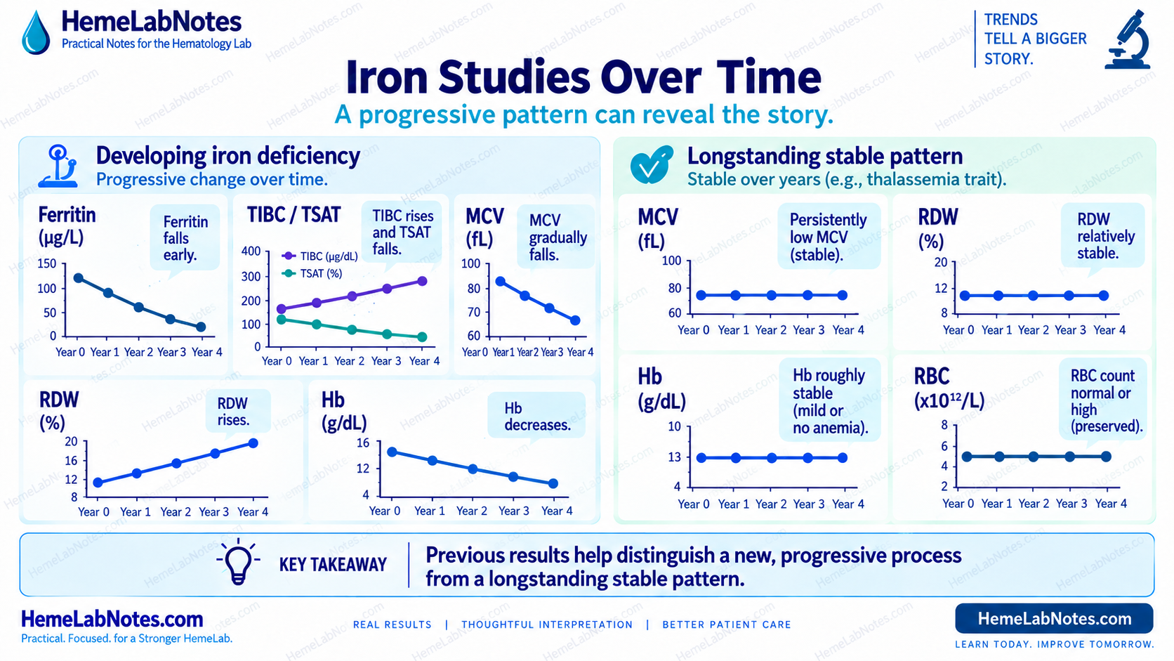
Iron studies are not exempt from trend interpretation.
Compare them with the patient’s CBC history.
Six months ago
- Hb normal
- MCV 86 fL
- RDW normal
Three months ago
- Hb slightly lower
- MCV 80 fL
- RDW beginning to rise
Today
- Hb reduced
- MCV 72 fL
- RDW clearly increased
- ferritin low
- TSAT low
The current ferritin matters.
But the previous CBCs tell us that the process was developing before the final pattern became obvious.
That is why an old CBC can sometimes answer a question that another new calculation cannot.
The laboratory story starts before today’s sample.
Iron Deficiency Does Not Have to Start With Microcytosis
Another common shortcut is:
Normal MCV = no iron deficiency.
That does not hold.
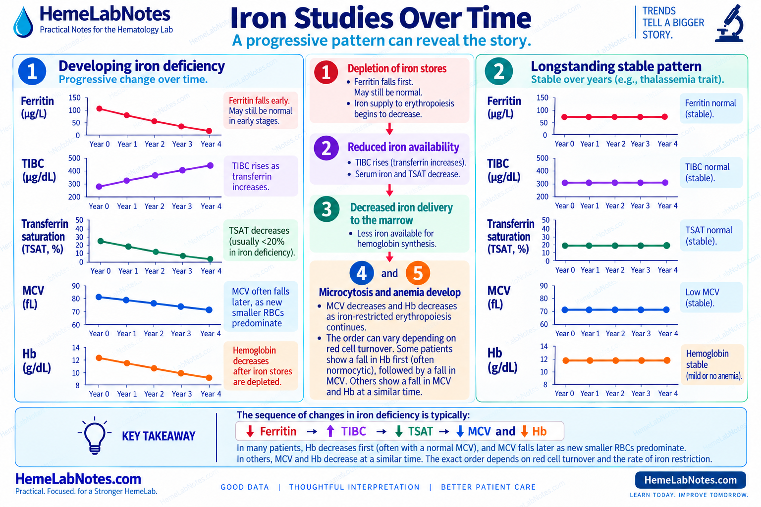
Iron stores may decline before the CBC becomes clearly microcytic.
Iron-deficient erythropoiesis can develop progressively, and MCV reduction may appear later in the process. [3]
A patient may therefore have:
- low ferritin
- falling TSAT
- normal MCV
especially earlier in iron depletion.
That is why iron studies should not be interpreted only after the MCV crosses a particular cutoff.
RDW Can Give an Earlier Hint
As iron availability becomes uneven, newly produced red cells may become progressively smaller while older cells remain in circulation.
The population becomes more variable.
RDW may rise.
The MCV may fall later.
The sequence can sometimes look like:
normal MCV → increasing RDW → falling MCV → worsening anemia
Not every patient follows that pattern.
The timing of Hb and MCV changes can vary with red-cell turnover and the pace of iron depletion.
But when the trend appears, it is useful.
Again:
trend before label.
What If Ferritin and TSAT Disagree?
This is where the panel earns its value.
Suppose ferritin looks acceptable but TSAT is low.
Before deciding what that means, ask:
- Is inflammation present?
- Is there chronic kidney disease?
- Is there liver disease?
- Is TIBC high, normal, or low?
- Does the CBC look iron-restricted?
- Are previous results moving?
- Has the patient recently received iron?
- Could more than one process be present?
Do not solve a discordant panel by choosing whichever result best fits the diagnosis you already expected.
Discordance is information.
It is a reason to slow down.
What About Soluble Transferrin Receptor?
Soluble transferrin receptor — sTfR — may sometimes help when ferritin is difficult to interpret, particularly when inflammation complicates the picture.
It tends to rise with iron-deficient erythropoiesis and is less directly influenced by inflammation than ferritin.
But it has limitations.
Increased erythropoietic activity can also raise it, and availability and assay interpretation vary between laboratories. [2,6]
So sTfR is not a replacement for the pattern.
It is another tool when the usual panel leaves the question open.
The sTfR/log ferritin index may also be useful as an adjunct in selected settings when distinguishing absolute iron deficiency from inflammatory iron restriction is difficult.
Again, this is a supporting tool — not a standalone diagnosis.
What About Reticulocyte Hemoglobin?
Reticulocyte hemoglobin measurements, reported as CHr or RET-He depending on the analyzer, can provide information about recent iron availability to erythropoiesis.
Because reticulocytes reflect more recent marrow production than mature circulating red cells, RET-He or CHr may sometimes help when conventional iron markers are difficult to interpret.
They can be particularly useful as an adjunct in mixed or inflammatory patterns.
But interpretation remains analyzer- and context-dependent.
Use the laboratory’s validated reference limits and clinical framework.
A Practical Bench Comparison
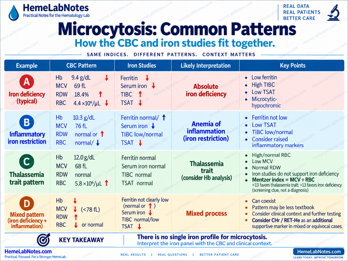
Example A — Straightforward iron depletion
- Hb: 9.4 g/dL
- MCV: 69 fL
- RBC count: 4.4 ×10⁶/µL
- RDW: 18.4%
- ferritin: low
- serum iron: low
- TIBC: high
- TSAT: low
The CBC and iron panel agree.
The next question is the cause of the iron deficiency.
Example B — Inflammatory iron restriction
- Hb: 10.3 g/dL
- MCV: 76 fL
- serum iron: low
- TSAT: low
- TIBC: low or normal
- ferritin: increased
- inflammatory context present
The iron is not readily available.
But this does not look like uncomplicated absolute iron depletion.
The clinical context changes the interpretation.
Example C — Microcytosis but iron studies do not fit
- Hb: 12.0 g/dL
- MCV: 68 fL
- RBC count: 5.8 ×10⁶/µL
- RDW: relatively stable
- ferritin: not reduced
- TSAT: not reduced
- previous pattern: longstanding
The Mentzer index is:
68 ÷ 5.8 ≈ 11.7
That supports a thalassemia-type pattern as a screening clue.
It does not establish the diagnosis.
Do not keep forcing iron deficiency.
Ask whether a thalassemia-type process fits better.
Example D — Mixed picture
- Hb: reduced
- MCV: reduced
- RDW: increased
- ferritin: not clearly low
- TSAT: low
- inflammation present
One number cannot settle this pattern.
Absolute iron deficiency and inflammatory iron restriction may coexist.
Depending on local availability, additional markers such as:
- sTfR
- sTfR/log ferritin index
- CHr / RET-He
may help refine the question.
Sometimes the most accurate laboratory conclusion is simply:
The iron pattern is mixed and requires further correlation.
A Practical Bench Sequence
When iron studies are ordered for microcytosis:
1. Start with the CBC pattern Why were iron studies ordered?
2. Look at ferritin Clearly low, indeterminate, or increased?
3. Check serum iron Useful only in context.
4. Check TIBC / transferrin Increased, normal, or reduced?
5. Review TSAT Is circulating iron availability reduced?
TSAT can be calculated as:
Serum iron ÷ TIBC × 100
A TSAT below roughly 20% is commonly regarded as reduced, although interpretation depends on clinical context and local practice.
6. Ask about inflammation Could ferritin be misleading?
7. Compare previous results New, progressive, or longstanding?
8. Revisit RBC count and RDW Does the CBC still fit the proposed explanation?
9. Consider additional testing when needed Depending on the unresolved pattern:
- sTfR
- sTfR/log ferritin index
- RET-He / CHr
- hemoglobin analysis
- other targeted investigations
10. Ask the final bench question
Not:
“Is the ferritin normal?”
But:
“Do the CBC, iron studies, and clinical context tell the same story?”
The Takeaway
Iron studies work best as a panel.
Ferritin is powerful.
But it is not infallible.
Serum iron can fall for more than one reason.
TIBC can help distinguish patterns.
TSAT tells us about available iron, not the cause of reduced availability.
Inflammation can change the meaning of several of those numbers.
And when the usual panel does not fit, additional markers may help — but they still belong inside the wider pattern.
So when microcytosis raises the possibility of iron deficiency:
Do not ask whether one iron result is abnormal. Ask whether the entire iron pattern fits the CBC.
Pattern first. Panel second. Diagnosis later.
References
-
World Health Organization. WHO Guideline on Use of Ferritin Concentrations to Assess Iron Status in Individuals and Populations. Geneva: World Health Organization; 2020.
-
Fertrin KY. Diagnosis and management of iron deficiency in chronic inflammatory conditions: is too little iron making your patient sick? Hematology Am Soc Hematol Educ Program. 2020;2020(1):478–486. doi:10.1182/hematology.2020000132.
-
Camaschella C. Iron deficiency. Blood. 2019;133(1):30–39.
-
Weiss G, Ganz T, Goodnough LT. Anemia of inflammation. Blood. 2019;133(1):40–50.
-
ARUP Consult. Microcytic Anemia Testing Algorithm. Updated February 2024.
-
ARUP Consult. Iron Deficiency Anemia and Anemia of Chronic Disease/Anemia of Inflammation Testing Algorithm. Updated January 2024.
Further Reading
- Bain BJ, Bates I, Laffan MA. Dacie and Lewis Practical Haematology. 12th ed. Elsevier; 2017.
- Keohane EM, Preston MM, Mirza KM, Walenga JM, eds. Rodak’s Hematology: Clinical Principles and Applications. 7th ed. Elsevier; 2025.