A low MCV tells us that the average red blood cell is small.
It does not tell us why.
That is where the RBC count becomes useful.
Two patients may have the same MCV but very different red-cell patterns.
For example:
- Patient A: MCV 70 fL, RBC 3.7 ×10⁶/µL
- Patient B: MCV 70 fL, RBC 5.8 ×10⁶/µL
Same MCV.
Different RBC count.
Different pattern.
Different next question.
A relatively high RBC count can strengthen a pattern. It cannot establish the diagnosis.
Start With the Pattern, Not the RBC Count
The RBC count only makes sense when it is read with the rest of the CBC.
For this HemeLabNotes series, the example MCV ranges are:
- Microcytic: <78 fL
- Normocytic: ≥78 fL to <98 fL
- Macrocytic: ≥98 fL
Always use the validated reference intervals from your own laboratory.
Once microcytosis is confirmed, I read the RBC count together with:
- hemoglobin
- hematocrit
- RDW
- MCH
- previous CBCs
- iron studies
- analyzer information
- blood-film findings when indicated
The RBC count is not the answer.
It is one part of the pattern.
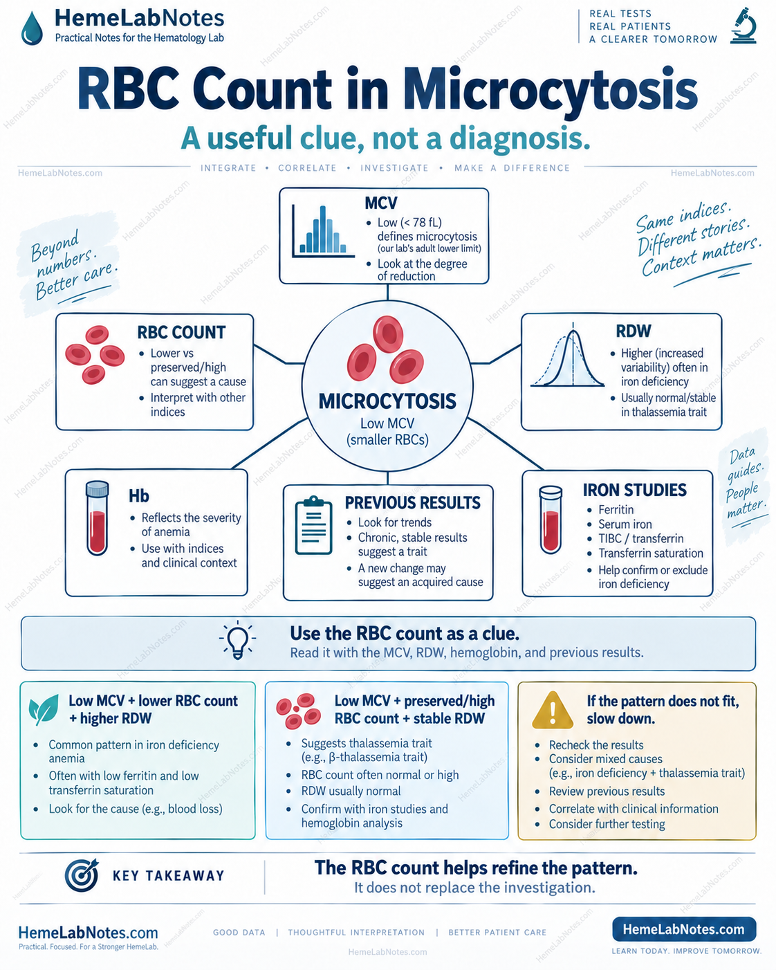
RBC count is one part of the broader microcytosis pattern. The previous article, Microcytic Anemia: How I Read the Pattern, outlines the full bench sequence.
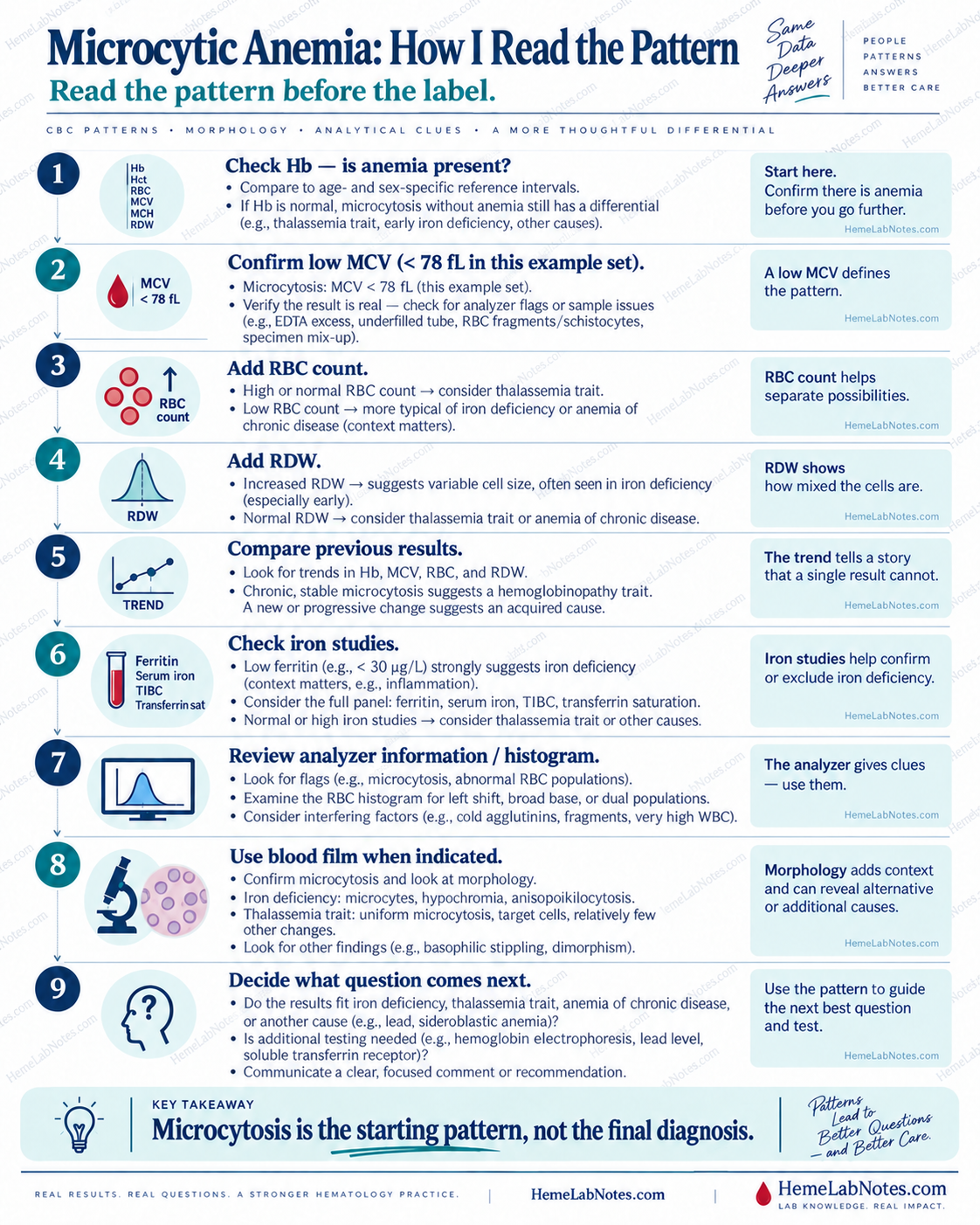
Why the RBC Count Matters Here
Microcytosis caused by iron restriction often behaves differently from a thalassemia-type pattern.
That does not mean the RBC count can diagnose either condition.
It means the count can change the direction of the next question.
Consider two simplified patterns.
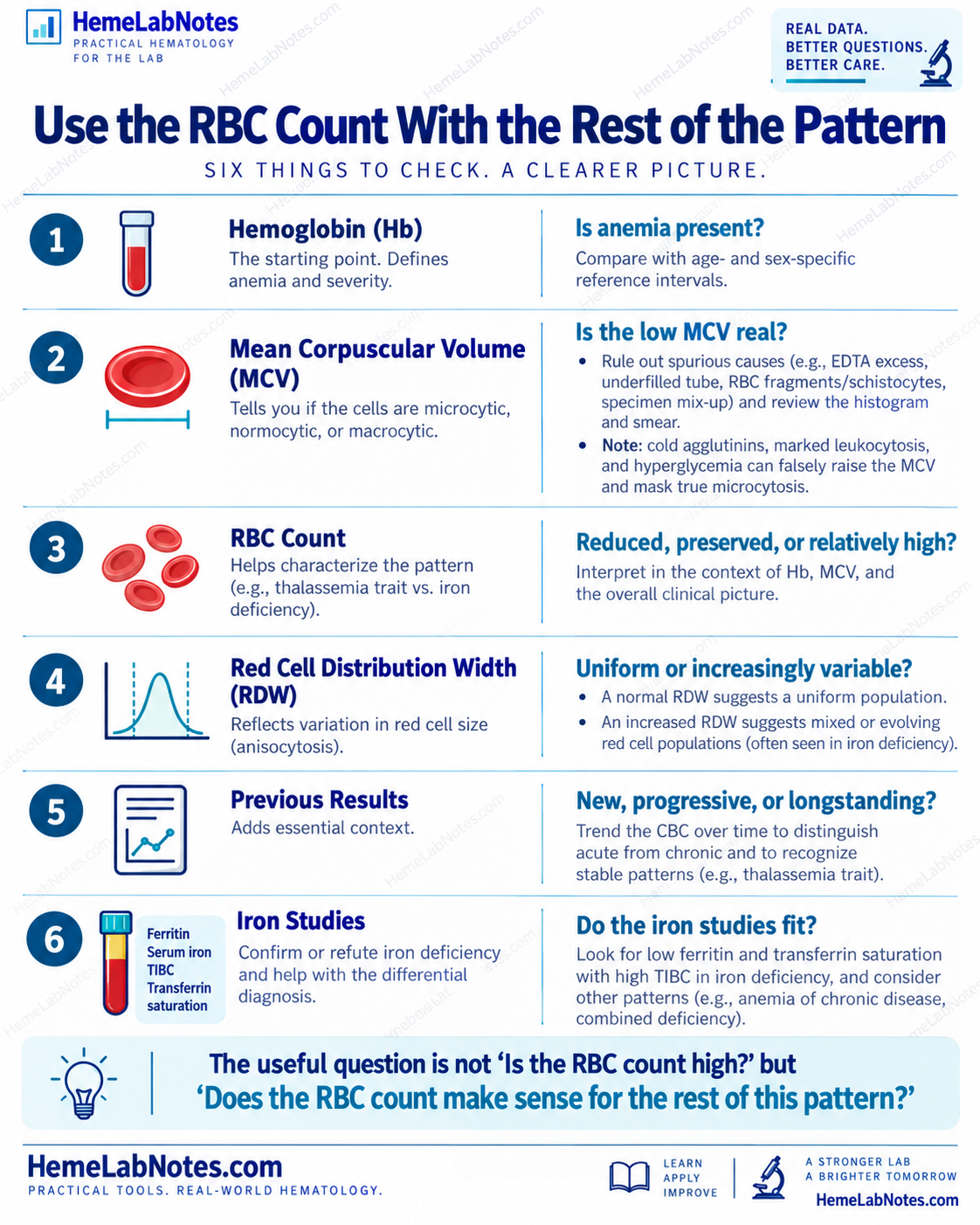
Pattern 1
- Hb: 10.2 g/dL
- MCV: 70 fL
- RDW: increased
- RBC count: 3.8 ×10⁶/µL
The RBC count is not preserved.
The RDW is increased.
This pattern may support an iron-restricted process, but iron studies are still needed.
Pattern 2
- Hb: near the lower reference interval
- MCV: 70 fL
- RDW: stable
- RBC count: 5.8 ×10⁶/µL
The RBC count is relatively preserved or high despite the low MCV.
That makes a thalassemia-type pattern worth considering.
It does not prove thalassemia.
Same MCV. Different RBC count. Different pattern.
A High RBC Count Does Not Automatically Mean Erythrocytosis
This is an important distinction.
A relatively high RBC count in microcytosis does not automatically mean that the patient has erythrocytosis.
Read it together with:
- hemoglobin
- hematocrit
- MCV
A patient may have many small red cells without having an increased red-cell mass.
That is why the RBC count should not be interpreted in isolation.
If the RBC count looks unusually high but the hemoglobin and hematocrit do not fit, slow down.
Ask whether the values are internally consistent.
Check Whether the Hb and Hct Fit
CBC parameters are related.
They should tell a reasonably coherent story.
If they do not, consider:
- specimen quality
- analytical interference
- abnormal morphology
- recent transfusion
- previous results
- analyzer flags
A discordant result is not something to force into a diagnosis.
It is a reason to verify.
If the pattern does not fit, verify before explaining it.
RDW Changes the Meaning of the RBC Count
The RBC count becomes more useful when RDW is added.
A preserved RBC count with longstanding microcytosis and a relatively stable RDW tells a different story from:
- preserved RBC count
- low MCV
- progressively widening RDW
- falling hemoglobin
The first may fit a longstanding inherited pattern.
The second may suggest that something new has been added.
For example, iron deficiency can develop in a patient who already has a thalassemia-type baseline.
So:
A chronic pattern does not protect the patient from a new problem.
Previous Results Often Beat a Single RBC Count
One of the most useful questions is:
Has this pattern always been here?
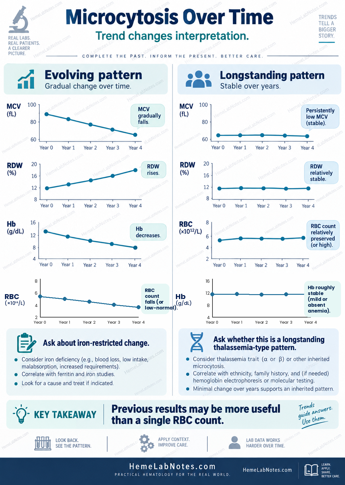
Imagine two patients with an MCV of 69 fL.
Patient A
Previous CBCs:
- MCV 86 fL
- then 80 fL
- then 76 fL
- now 69 fL
Hemoglobin is falling.
RDW is increasing.
This is an evolving pattern.
Patient B
Previous CBCs over several years:
- MCV 68–70 fL
- RBC count 5.6–5.9 ×10⁶/µL
- RDW relatively stable
- hemoglobin roughly stable
This is a longstanding pattern.
The current MCV is similar.
The story is not.
The number matters. The change in the number often matters more.
Iron Deficiency and a Thalassemia-Type Pattern Can Coexist
Real patients do not always fit one clean category.
A patient may have longstanding microcytosis with a relatively preserved RBC count and later develop iron deficiency.
That can change the pattern.
You may see:
- MCV fall further
- hemoglobin decrease
- RDW increase
- RBC count become less preserved
- iron studies become abnormal
This is why a familiar longstanding CBC pattern should not end the investigation when something has changed.
Mixed conditions make simple rules less reliable.
What About the Mentzer Index?
The Mentzer index is:
MCV ÷ RBC count
It was developed as a screening aid for differentiating iron-deficiency-type patterns from thalassemia-type patterns.
A commonly used interpretation is:
- <13: favors a thalassemia-type pattern
-
13: favors iron deficiency
For example:
MCV 72 fL
RBC count 4.4 ×10⁶/µL
Mentzer index:
72 ÷ 4.4 ≈ 16.4
That may support an iron-deficiency-type pattern.
But the Mentzer index is not a diagnosis.
Its performance varies between populations, and mixed conditions reduce its usefulness.
So I use it as another clue.
Not as the final answer.
Iron Studies Still Matter
If the CBC suggests iron restriction, move to the iron panel.
That may include:
- ferritin
- serum iron
- TIBC or transferrin
- transferrin saturation
A low ferritin strongly supports depleted iron stores.
But ferritin is also an acute-phase reactant.
Inflammation can raise ferritin, so a normal or elevated result does not automatically exclude iron deficiency.
That is why the iron panel needs clinical context.
Again:
The pattern matters more than one number.
When the RBC Count Misleads
The RBC count is useful, but there are situations where it becomes harder to interpret.
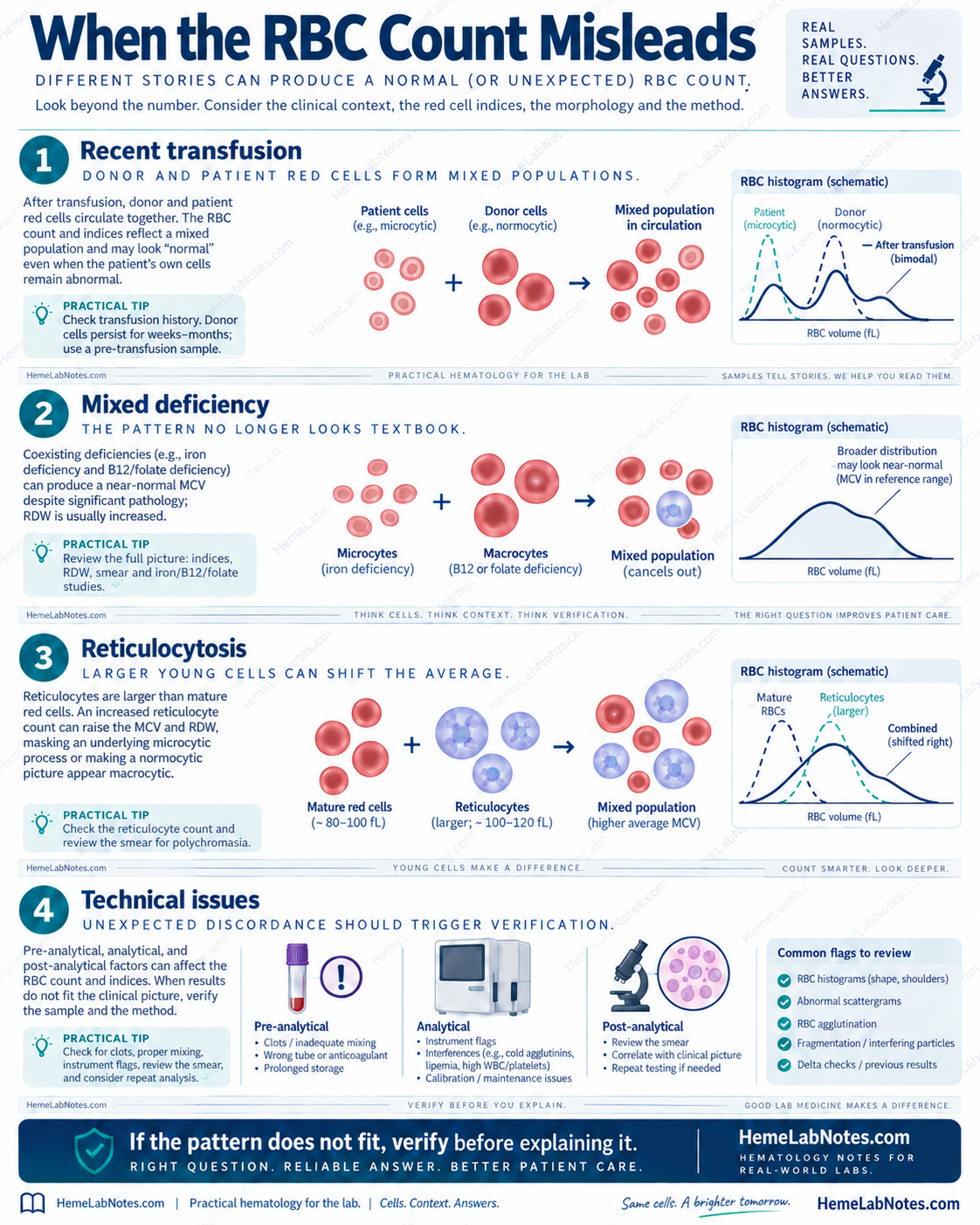
Recent transfusion
Donor and patient red cells circulate together.
The CBC may represent a mixed population rather than the patient’s original pattern.
Mixed deficiency
Iron deficiency and B12 or folate deficiency may pull the indices in different directions.
The average may look less abnormal than the underlying biology.
Reticulocytosis
Reticulocytes are larger than mature red cells.
An increased reticulocyte population can shift the MCV and RDW.
Technical issues
Pre-analytical and analytical problems can also create unexpected discordance.
Review:
- sample quality
- analyzer flags
- histograms
- scattergrams
- blood film
- delta checks
- previous results
The RBC count is useful only when the result itself is trustworthy.
Analyzer Information Can Add Context
Do not ignore the analyzer.
Look at:
- RBC histogram
- distribution width
- left shift
- broadening
- shoulders
- dual populations
- abnormal flags
A left-shifted RBC histogram supports the presence of smaller cells.
A broadened distribution may support anisocytosis.
A mixed or bimodal population may suggest more than one red-cell population.
Interference can also alter the apparent pattern.
For example:
- cold agglutinins can decrease the measured RBC count and falsely increase MCV
- marked leukocytosis can interfere with some CBC measurements
- severe hyperglycemia may increase MCV without changing the RBC count
These do not create true microcytosis.
They can make the pattern harder to interpret.
Use the Blood Film When Indicated
A blood film can add information that the indices cannot.
Look for:
- microcytosis
- hypochromia
- anisocytosis
- poikilocytosis
- target cells
- elongated or pencil-like cells
- mixed red-cell populations
- other unexpected abnormalities
No single morphology is diagnostic.
Target cells are not exclusive to thalassemia.
Pencil cells are not exclusive to iron deficiency.
Use morphology to strengthen or challenge the pattern.
Not to replace the rest of the workup.
Three Practical Examples
Example A — Iron-restricted pattern
- Hb: 9.8 g/dL
- MCV: 69 fL
- RBC count: 3.7 ×10⁶/µL
- RDW: 18.5%
- previous results: MCV and Hb falling
This pattern makes iron studies an appropriate next step.
The RBC count supports the question.
It does not answer it.
Example B — Longstanding thalassemia-type pattern
- Hb: 12.1 g/dL
- MCV: 69 fL
- RBC count: 5.8 ×10⁶/µL
- RDW: 13%
- previous pattern: stable for years
This is a different pattern.
The relatively preserved RBC count and stable RDW make a thalassemia-type process worth considering.
Iron studies should still be reviewed.
Hemoglobin analysis may be appropriate depending on the clinical context.
Example C — The pattern is not neat
- Hb: 10.5 g/dL
- MCV: 66 fL
- RBC count: 5.4 ×10⁶/µL
- RDW: 19%
The RBC count is preserved.
But the RDW is increased.
That does not fit a simple textbook pattern.
Do not force it.
Check:
- previous CBCs
- iron studies
- possible mixed causes
- hemoglobin analysis when appropriate
- analyzer information
- blood film
A pattern that does not fit is a reason to slow down, not a reason to force an answer onto it.
A Practical Bench Sequence
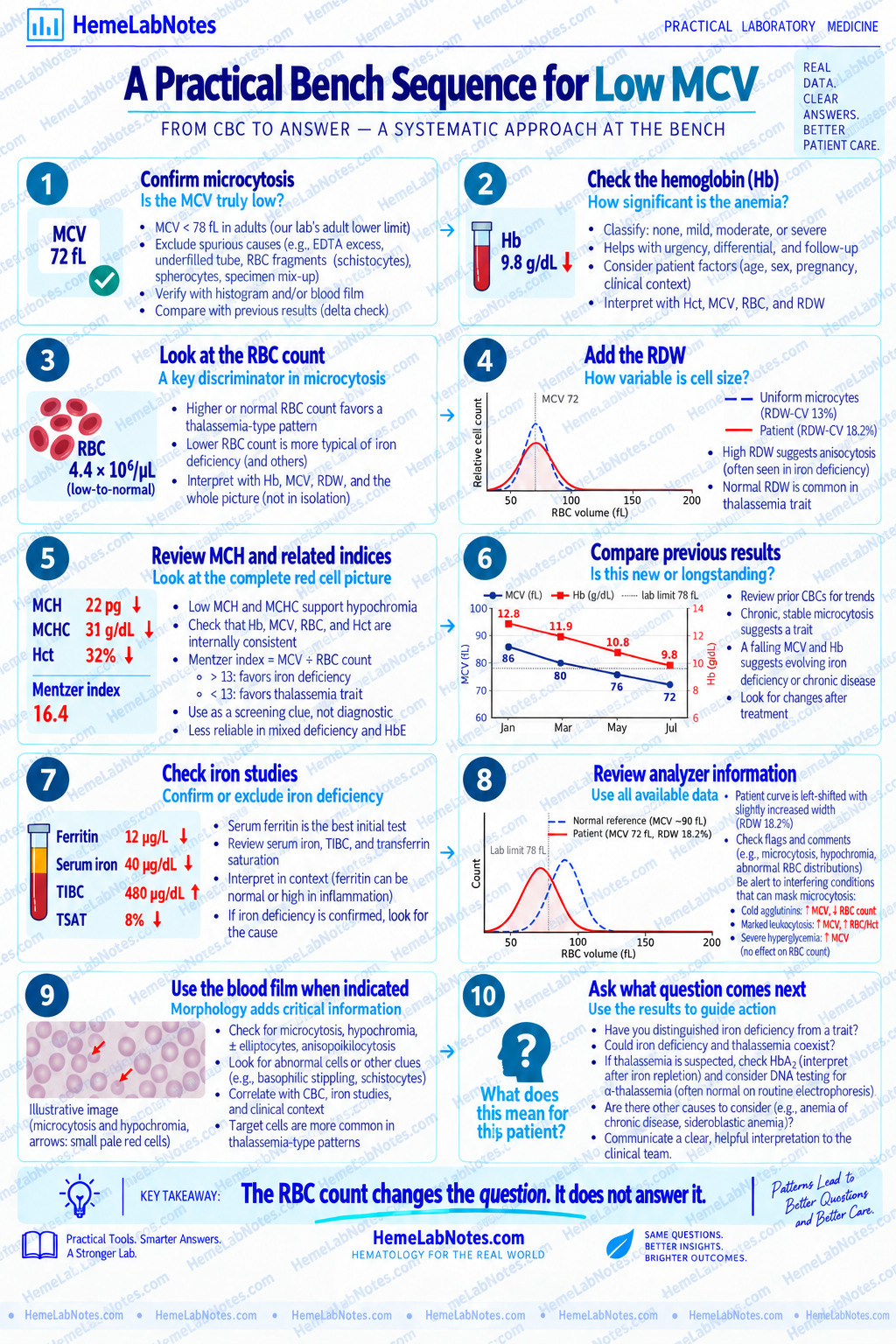
When I see a low MCV, I work through the pattern in this order:
1. Confirm the MCV
Is the microcytosis real?
2. Check the hemoglobin
Is anemia present, and how significant is it?
3. Look at the RBC count
Reduced, preserved, or relatively high?
4. Add the RDW
Uniform or increasingly variable?
5. Review MCH and related indices
Does the red-cell picture remain internally consistent?
6. Compare previous results
New, progressive, or longstanding?
7. Check iron studies
Do they support iron deficiency?
8. Review analyzer information
Do the histogram and flags fit?
9. Use the blood film when indicated
Does morphology add or challenge the interpretation?
10. Ask what question comes next
Iron deficiency?
Thalassemia-type pattern?
Mixed process?
Another cause?
The final question is not:
“Is the RBC count high?”
It is:
“Does the RBC count make sense for the rest of this pattern?”
The Takeaway
The RBC count can sharpen the interpretation of microcytosis.
It can help distinguish one pattern from another.
It can make a thalassemia-type process more plausible.
It can make an iron-restricted process more plausible.
But it cannot replace:
- iron studies
- previous results
- analyzer information
- morphology
- clinical context
- appropriate confirmatory testing
The RBC count changes the question. It does not answer it.
Read the pattern.
Then decide what question comes next.
References
-
Bain BJ, Bates I, Laffan MA. Dacie and Lewis Practical Haematology. 12th ed. Elsevier; 2017.
-
Bain BJ. Blood Cells: A Practical Guide. Wiley-Blackwell.
-
Keohane EM, Preston MM, Mirza KM, Walenga JM, eds. Rodak’s Hematology: Clinical Principles and Applications. Elsevier.
-
Camaschella C. Iron-deficiency anemia. N Engl J Med. 2015;372(19):1832–1843. doi:10.1056/NEJMra1401038.
-
Mentzer WC Jr. Differentiation of iron deficiency from thalassaemia trait. Lancet. 1973;1(7808):882. doi:10.1016/S0140-6736(73)91446-3.
-
ARUP Consult. Microcytic Anemia Testing Algorithm. Updated February 2024.
-
Merck Manual Professional Edition. Evaluation of Anemia.