Sometimes a CBC looks wrong before you even look at the blood film.
The RBC count is unexpectedly low. The MCV is high, and the MCHC is higher than we would normally expect. Yet the haemoglobin does not seem to fit the rest of the picture.
This is where I stop looking for a diagnosis and start asking whether the analyser has measured the sample correctly.
One possibility is cold agglutination.
Recognising the pattern is often straightforward. The real challenge is deciding which results are affected, whether the interference has been adequately corrected, and what can safely be reported.
1. When the CBC Stops Making Sense
The first clue is usually not one abnormal parameter. It is a group of results that no longer agree.
In a typical sample, haemoglobin, haematocrit and RBC indices should form a reasonably consistent picture.
The familiar rule of three provides a quick check:
HGB (g/dL) × 3 ≈ Hct (%)
This is only an approximation, not a formal acceptance criterion. Nevertheless, a large discrepancy deserves investigation.
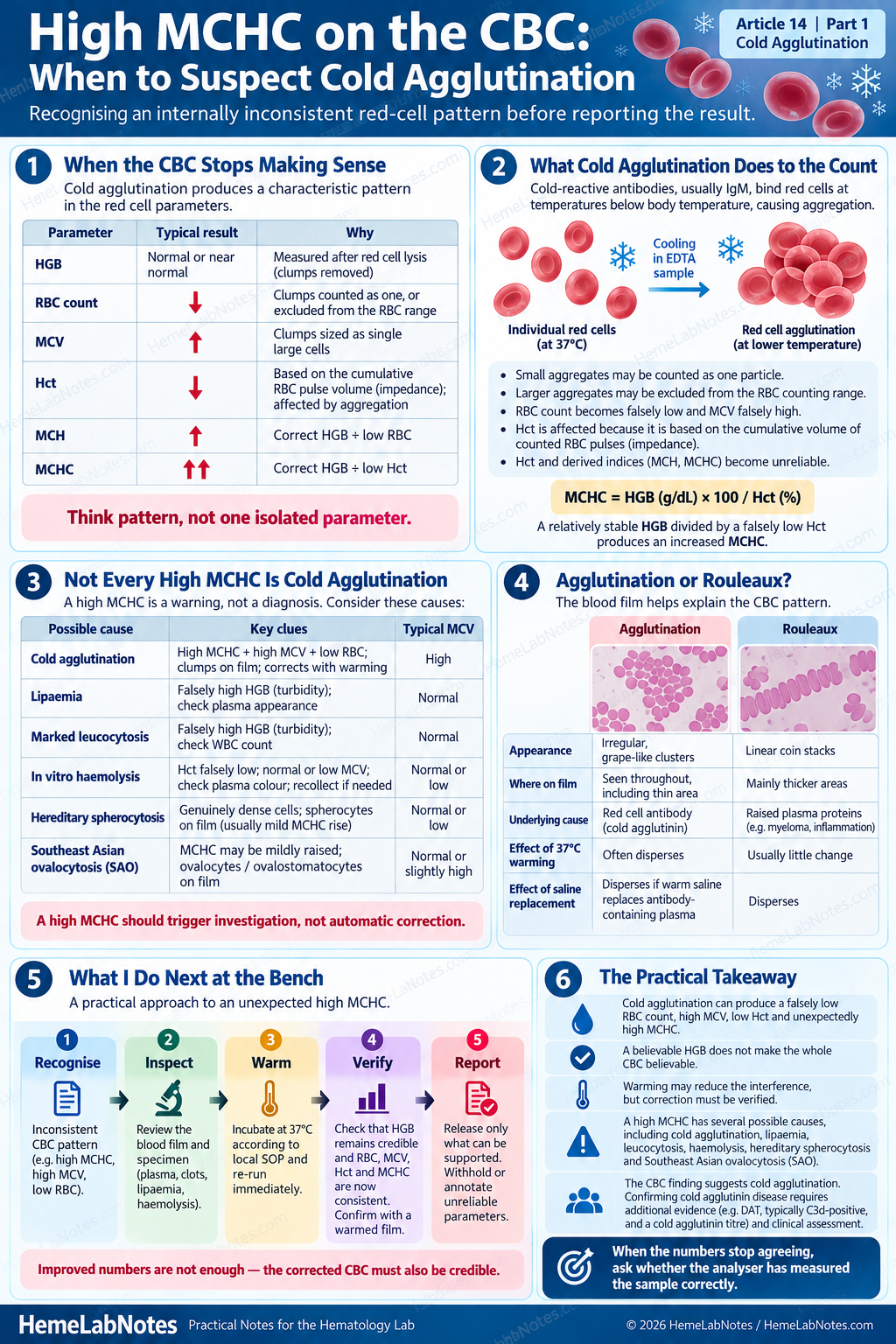
Cold agglutination can produce a characteristic pattern:
| Parameter | Typical change | Explanation |
|---|---|---|
| HGB | Often relatively unchanged | RBCs are lysed before photometric measurement |
| RBC | Falsely decreased | Aggregates counted as one particle or excluded |
| MCV | Falsely increased | Aggregates interfere with RBC sizing |
| Hct | Often falsely decreased | Cumulative RBC volume becomes unreliable |
| MCH | Falsely increased | HGB divided by underestimated RBC |
| MCHC | Falsely increased | HGB divided by underestimated Hct |
These changes begin when cold-reactive antibodies, usually IgM, cause red cells to aggregate as the specimen cools.
The degree of aggregation depends on factors including antibody concentration, thermal amplitude and specimen temperature.
Why the analyser gets confused
During impedance measurement, red cells pass through an aperture and generate electrical pulses.
The analyser expects each event to represent an individual cell.
But what happens when two or three red cells are stuck together?
A small aggregate may be registered as a single, unusually large particle. Larger aggregates may fall outside the accepted RBC measurement range.
The result is an underestimated RBC count and a distorted red-cell volume distribution.
The errors then affect calculated parameters.
MCH and MCHC may rise to implausible values even though the patient’s individual red cells are not genuinely hyperchromic.
One distinction matters here.
Conventional HGB is usually less affected by cold agglutination itself, because the red cells are lysed before measurement.
However, HGB is not automatically reliable if another interference, such as lipaemia or substantial plasma haemolysis, is also present.
A believable HGB does not make the entire CBC believable.
2. Why MCHC Is an Important Warning
MCHC is one of the most useful indicators of an unreliable CBC.
It is calculated as:
MCHC (g/dL) = HGB (g/dL) × 100 / Hct (%)
If HGB remains relatively stable while Hct is falsely reduced, the calculated MCHC increases.
Values above approximately 36–37 g/dL often trigger investigation, although the appropriate review threshold depends on the analyser and the laboratory’s validated rules. [1,2]
But a high MCHC does not automatically mean cold agglutination.
There are other possibilities.
| Possible cause | Main problem | Useful check |
|---|---|---|
| Cold agglutination | RBC counting and volume interference | Blood film, warming |
| Lipaemia | Photometric HGB interference | Plasma appearance, validated correction |
| Marked leucocytosis | Possible HGB or counting interference | WBC count, scattergrams |
| In-vitro haemolysis | Loss of intact RBCs with plasma-free haemoglobin | Plasma inspection |
| Hereditary spherocytosis | Genuinely increased cellular haemoglobin concentration may occur | Blood film, further investigations |
| Southeast Asian ovalocytosis (SAO) | MCHC may be mildly raised | Ovalocytes / ovalostomatocytes on blood film [3] |
The MCV often provides another clue.
A high MCHC accompanied by unexpectedly high MCV and reduced RBC count should raise suspicion of agglutination.
A high MCHC with relatively stable RBC indices may suggest a different mechanism.
However, these are patterns, not diagnostic rules.
This is why I would not stop at the numbers.
The next step is to examine the blood film.
3. Agglutination or Rouleaux? The Blood Film Matters
Cold agglutination produces irregular red-cell clusters.
The cells do not follow a consistent orientation, and aggregates may remain visible even in relatively well-spread areas of the blood film.
Sometimes the aggregates are striking.
Other times, the findings are subtle enough that a quick glance may miss them.
One important distinction is between agglutination and rouleaux.
Rouleaux forms linear stacks of red cells resembling coins. It is often associated with increased plasma proteins, including inflammatory conditions and plasma-cell disorders.
| Feature | Agglutination | Rouleaux |
|---|---|---|
| Arrangement | Irregular clusters | Linear stacks |
| Main mechanism | Antibody-mediated RBC aggregation | Plasma-protein-related stacking |
| Blood film | May persist in thin areas | Often more prominent in thicker areas |
| Warming to 37°C | May disperse cold-reactive aggregates | Usually little effect |
A saline-dispersion technique can help distinguish rouleaux from agglutination, although the method and temperature must be considered when cold-reactive antibodies are involved.
For a CBC affected by suspected cold agglutination, I find another comparison particularly useful: examine the original film alongside a film prepared after controlled warming.
If the red-cell clusters disperse and the RBC indices improve together, the findings strongly support temperature-dependent agglutination.
But there is an important limitation.
Seeing red-cell agglutination is not the same as diagnosing cold agglutinin disease.
The blood film identifies an abnormal laboratory phenomenon. Establishing the underlying clinical condition requires additional evidence.
Where clinically indicated, investigations may include a direct antiglobulin test (DAT), typically showing C3d reactivity in cold agglutinin disease, together with appropriately performed cold agglutinin titre testing and assessment for haemolysis. [4]
A positive C3d DAT alone does not establish the diagnosis.
For the immediate CBC investigation, our concern is whether the aggregation has interfered with measurement.
4. Warming the Sample: Correction or Just Improvement?
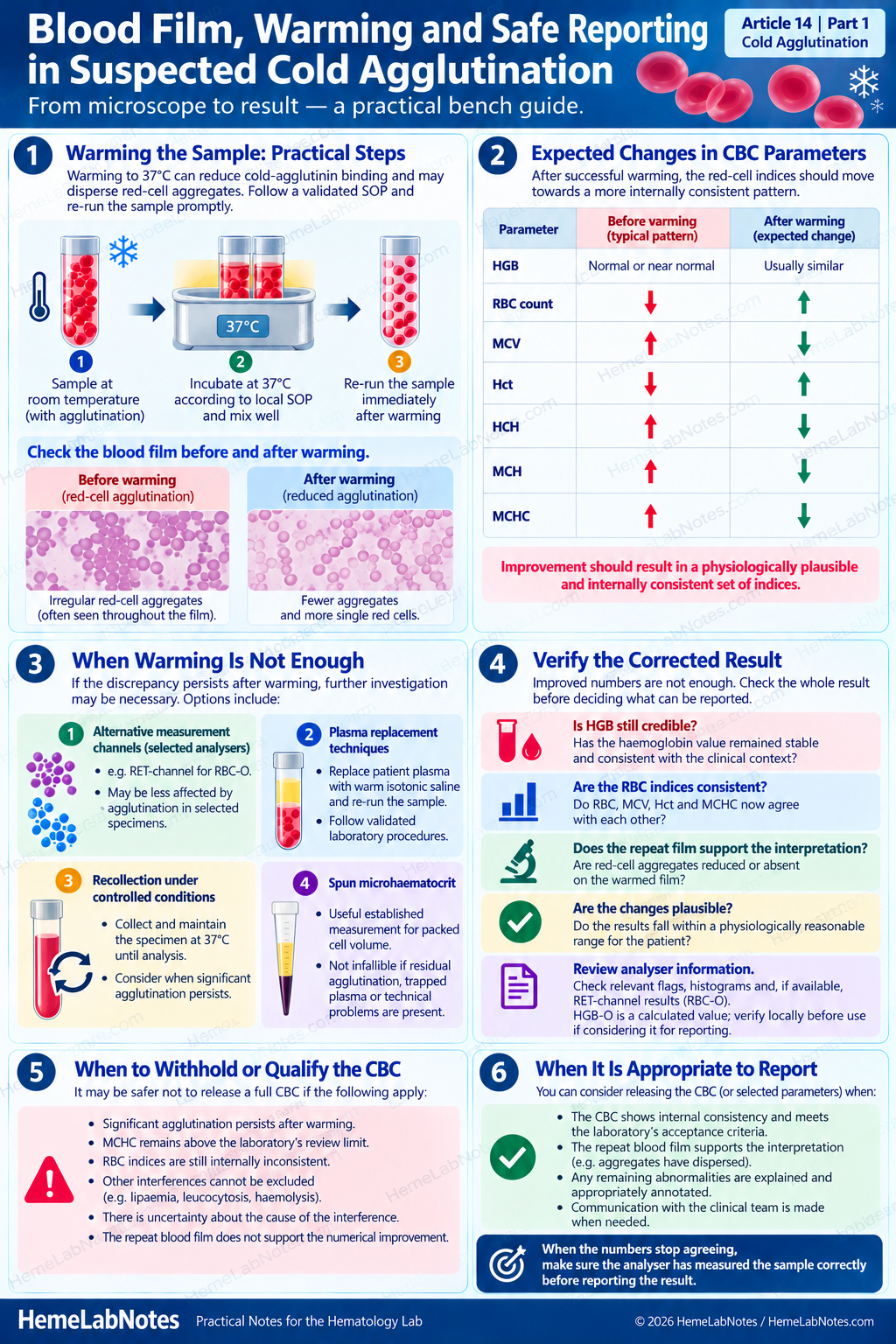
Warming the specimen to 37°C remains a conventional method for reducing cold-agglutinin interference.
As the temperature rises, cold-reactive antibody binding may decrease and the aggregates may disperse.
The specimen is then mixed and analysed promptly according to the laboratory’s validated SOP.
The timing matters because agglutination may return as the sample cools.
After successful warming, we would generally expect:
- RBC count to increase towards a more credible value
- MCV to decrease
- Hct to become more consistent with HGB
- MCH and MCHC to move towards physiologically plausible values
- Red-cell aggregates to diminish or disappear on the repeat film
HGB should usually change relatively little when cold agglutination is the principal interference.
But this is where I would be careful.
An improved CBC is not necessarily a corrected CBC.
Suppose the MCHC falls considerably after warming but remains above the laboratory’s review limit.
Does that mean the specimen is now acceptable?
Not necessarily.
Residual agglutination may still affect RBC counting and the derived indices.
Does longer warming always produce better results?
Not necessarily.
La Gioia and colleagues compared several approaches to reducing cold-agglutinin interference on Sysmex analysers. [5]
They found that preheating at 41°C for one minute produced reversal comparable to the RET-channel approach and better results than incubation at 37°C for two hours under their study conditions.
That is an interesting finding.
It suggests that a longer incubation at a lower temperature does not necessarily provide better analytical correction.
However, the authors also concluded that none of the investigated procedures guaranteed complete reversal of agglutination in every specimen.
The result should not be interpreted as a recommendation to replace a validated 37°C SOP with routine 41°C warming.
It shows why the effectiveness of a correction method must be demonstrated rather than assumed.
What if warming does not resolve the discrepancy?
Further investigation may be necessary.
Depending on the suspected interference and the laboratory’s validated procedures, options may include alternative measurement channels, appropriate plasma-replacement techniques, recollection under controlled temperature conditions or selected manual measurements.
A spun microhaematocrit remains a useful established measurement and underpins reference approaches to packed cell volume. However, it is not infallible when residual aggregation, trapped plasma or technical problems are present.
Its suitability should be assessed in the context of the specimen and method.
When reliable RBC indices cannot be established, the laboratory may need to withhold or qualify the affected parameters while reporting measurements that can be supported.
Releasing a complete CBC is not more important than releasing an accurate one.
5. A Practical Approach at the Bench
When an unexpectedly high MCHC appears, I prefer to work through the problem in a consistent sequence.
First, recognise the inconsistency.
Review HGB, RBC, Hct, MCV, MCH and MCHC together. Check relevant analyser flags and previous patient results.
Does the pattern suggest an RBC counting problem, a haemoglobin interference or a genuine red-cell abnormality?
Next, investigate the specimen.
Inspect the plasma when appropriate and review the peripheral blood film.
Look for irregular red-cell aggregates, rouleaux, spherocytes, ovalostomatocytes or other abnormalities that might explain the indices.
Then, apply the appropriate corrective method.
For suspected cold agglutination, this may involve controlled warming and repeat analysis.
Other causes of high MCHC require different approaches.
Then, verify the whole result.
Has HGB remained credible, and are RBC, MCV, Hct and MCHC now consistent with each other? Does the repeat film support the interpretation?
Finally, decide what can be reported.
Check that the selected results satisfy the laboratory’s analytical acceptance and reporting criteria.
If uncertainty remains, affected parameters should be withheld or appropriately annotated, with clinical communication when necessary.
The aim is not to force every parameter into the reference interval.
It is to establish which values reflect the patient’s blood rather than the analytical interference.
6. Beyond Warming: The Role of Optical RBC Measurements
Traditional corrective methods can take time, especially when the sample requires incubation, repeat analysis and additional film review.
Modern haematology analysers provide another possibility.
Published investigations on Sysmex XE and XN platforms demonstrated that optical RBC counting in the reticulocyte (RET) channel may be less affected by cold agglutination in selected specimens. [1,5]
The RET channel uses fluorescence flow cytometry and different reagent and thermal conditions from conventional impedance counting.
On the XN-10 studied by Berda-Haddad and colleagues, the RET channel provided a second RBC measurement, known as RBC-O, following reagent treatment and internal warming. [1]
The researchers also described HGB-O, an alternative haemoglobin estimate calculated mainly from RBC-O and RBC haemoglobin-equivalent information (RBC-He).
This distinction is important.
RBC-O is a measured optical count. HGB-O is a calculated haemoglobin estimate, not a second conventional photometric HGB measurement.
The two parameters do not necessarily have the same analytical limitations.
What about the newer Sysmex XR-Series?
The XR-Series uses related analytical technologies, and appropriately configured instruments offer an optional RET channel.
Sysmex also describes CBC-O as an optional Extended IPU workflow optimiser designed to investigate increased MCHC and suggest alternative reportable parameters. [6]
However, the original published cold-agglutination studies discussed here were performed on earlier XE/XN systems.
The availability and intended operation of RET and CBC-O on XR systems are supported by manufacturer documentation. That does not, by itself, establish equivalent analytical performance for every XR configuration or specimen.
This creates an interesting question:
Can optical measurements provide a reliable CBC without a separate manual warming step?
Possibly, but there are limitations.
A measured optical RBC count, a derived HGB-O value and recalculated red-cell indices do not all carry the same analytical assumptions.
A CBC that appears internally consistent is not automatically analytically accurate.
That is why alternative measurements still require appropriate verification before replacing conventional results.
The principle is promising.
The more useful question is how well it performs when faced with an actual problematic specimen.
The Takeaway
Cold agglutination can make a CBC internally inconsistent.
A falsely low RBC count, increased MCV, reduced Hct and unexpectedly high MCHC may all result from red-cell aggregation rather than genuine changes in red-cell size or haemoglobin concentration.
The blood film helps identify the problem, and warming often reduces the interference.
But correction should not be judged by numerical improvement alone.
Before releasing the CBC, we need to establish that the reported measurements are sufficiently reliable.
When the numbers stop agreeing, the first question is not what disease the patient has. It is whether the analyser has measured the sample correctly.
Coming Next — Part 2 (Article 15)
Cold Agglutination: Can CBC-O Resolve the Interference Without Manual Warming?
In Part 2, we will examine a laboratory case using the Sysmex XR-Series, compare conventional CBC and optical measurements, and explore how CBC-O parameters can help guide reporting decisions.
The focus will be on what the analyser actually measures, what it calculates, and whether the alternative results can be trusted.
References
1. Berda-Haddad Y, Faure C, Boubaya M, Arpin M, Cointe S, Frankel D, Lacroix R, Dignat-George F. Increased mean corpuscular haemoglobin concentration: artefact or pathological condition? International Journal of Laboratory Hematology. 2017;39(1):32–41.
https://doi.org/10.1111/ijlh.12565
2. Girard S, Berda-Haddad Y, Brouzes C, et al. Breaking Free From MCHC Interferences? French-Speaking Cellular Haematology Group (GFHC) Review of Causes, Rising Trends and Practical Solutions. International Journal of Laboratory Hematology. 2025;47(5):798–807.
https://doi.org/10.1111/ijlh.14536
3. Souissi M, Daliphard S, Picard V, Lebigot E, Jardin F, Bobée V. Elevated MCHC reveals a Southeast Asian Ovalocytosis. American Journal of Hematology. 2024;99(2):331–332.
https://doi.org/10.1002/ajh.27157
4. Ubezio G, Barcellini W, Coluzzi S, Fattizzo B, Matteocci A, Patriarca A, et al. Standardized diagnostic approach for cold agglutinin disease: results from a Delphi-Based Expert Consensus. Blood Transfusion. Published online July 15, 2026.
https://doi.org/10.2450/BloodTransfus.1314
5. La Gioia A, Fumi M, Fiorini F, et al. Short preheating at 41°C leads to a red blood cells count comparable to that in RET channel of Sysmex analysers in samples showing cold agglutination. Journal of Clinical Pathology. 2018;71(8):729–734.
https://doi.org/10.1136/jclinpath-2017-204954
6. Sysmex. XR-Series Workflow Automation: CBC-O and Extended IPU. Product and workflow documentation.
https://www.sysmex-mea.com/products/diagnostics/haematology/xr-series-workflow-automation/
HemeLabNotes provides practical educational material for laboratory professionals. Interpretation, corrective procedures and reporting decisions should follow the laboratory’s validated methods, applicable standards and clinical context.