Sometimes the CBC looks familiar.
The MCV and MCH are low, the RBC count is relatively preserved, and the blood film shows the usual features of microcytosis and hypochromia.
At first glance, it looks like another thalassemia-trait pattern.
But something does not quite fit.
The hemoglobin is lower than expected. The blood film shows more anisopoikilocytosis, and the reticulocyte count is increased. There may even be a history of jaundice or episodes of worsening anemia.
This is where I start asking a different question.
Could this be more than alpha-thalassemia trait?
One possibility is hemoglobin H (HbH) disease.
Unlike an uncomplicated carrier state, HbH disease can produce enough alpha-globin deficiency to form abnormal β-globin tetramers, leading to chronic hemolysis and clinically significant anemia.
The severity varies considerably. Some patients remain relatively stable and are diagnosed only in adulthood. Others develop more substantial anemia, recurrent hemolytic episodes, or transfusion requirements.
The laboratory challenge is not simply recognizing microcytosis. It is recognizing when the wider pattern no longer fits a relatively stable carrier phenotype.
When the CBC Looks More Than a Trait
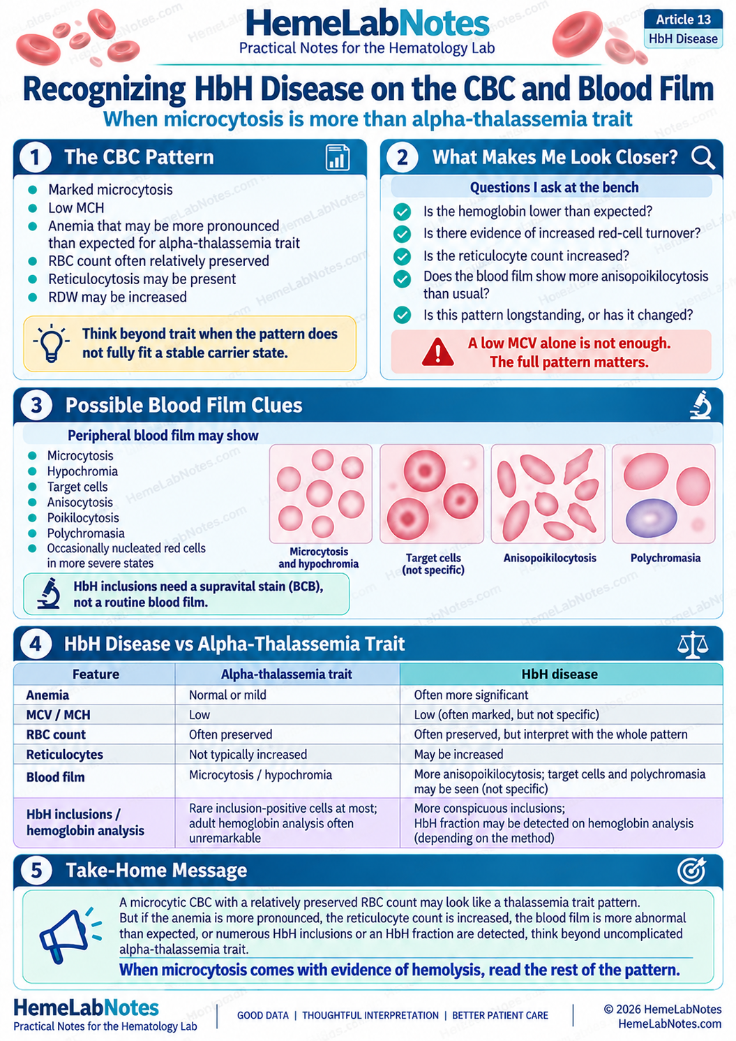
Alpha-thalassemia trait usually produces a relatively stable microcytic and hypochromic phenotype. Hemoglobin may be normal or mildly reduced, the RBC count is often preserved, and routine adult hemoglobin analysis may be unremarkable.
HbH disease belongs to a more clinically significant part of the alpha-thalassemia spectrum.
GeneReviews describes HbH disease as a microcytic, hypochromic hemolytic anemia with variable clinical severity. [1]
But I would not suspect HbH disease simply because the MCV is particularly low.
What interests me is what accompanies that microcytosis.
Is the hemoglobin disproportionately reduced? Is there increased red-cell turnover? Does the morphology suggest more than an uncomplicated carrier state? Have previous CBCs shown a relatively stable phenotype, or is something changing?
An increased reticulocyte count or a history of unexplained worsening anemia may provide additional clues.
No single finding establishes the diagnosis. But when several results point in the same direction, the possibility deserves investigation.
A longstanding microcytic phenotype with evidence of hemolysis is not the same laboratory problem as uncomplicated alpha-thalassemia trait.
What Actually Happens in HbH Disease
The mechanism begins with inadequate alpha-globin chain production.
Normally, alpha chains combine with beta chains to produce adult hemoglobin A:
HbA = α₂β₂
When alpha-chain production is significantly reduced, fewer normal HbA molecules can be assembled.
In classic deletional HbH disease, three of the four alpha-globin genes are deleted.
A typical genotype is:
--/-α
Only one functional alpha-globin gene remains.
The reduced alpha-chain production creates an imbalance, leaving excess beta-globin chains that associate to form tetramers:
HbH = β₄
These β₄ tetramers have an abnormally high oxygen affinity, making them less effective at releasing oxygen to tissues.
They are also unstable. Their precipitation within red cells contributes to membrane damage and premature red-cell destruction. Ineffective erythropoiesis may contribute to the anemia as well. [1,2]
The laboratory consequences include microcytosis, hypochromia, chronic hemolysis, increased erythropoietic activity, and anemia of varying severity.
That explains why HbH disease may produce a more complicated hematologic picture than uncomplicated alpha-thalassemia trait.
The red cells are not simply small. Their survival may also be shortened.
Reading the CBC and Reticulocyte Count Together
The CBC remains my starting point, but I would be cautious about placing too much weight on any single parameter.
Hemoglobin and RBC Count
HbH disease may cause mild, moderate, or occasionally severe anemia.
Many patients with deletional HbH disease maintain relatively stable hemoglobin concentrations without regular transfusions. Others have more substantial anemia, particularly with certain non-deletional variants. [3,4]
The RBC count may remain relatively preserved despite pronounced microcytosis.
This creates an important interpretation trap.
A relatively high RBC count may suggest a thalassemia-type phenotype, but it does not prove that the patient has only a clinically mild carrier state.
The degree of anemia and evidence of hemolysis still matter.
MCV, MCH, and RDW
MCV and MCH are usually reduced, sometimes markedly.
However, severe microcytosis is not specific to HbH disease. Iron deficiency and other thalassemia syndromes can produce similarly low indices.
RDW may be increased, reflecting greater variation in red-cell size, although it is not consistently abnormal in every case.
I would use these parameters to understand the overall pattern, not as independent diagnostic cutoffs.
Reticulocyte Count
This is where the interpretation becomes more interesting.
Because HbH disease involves chronic hemolysis, the bone marrow may respond by increasing red-cell production.
GeneReviews describes moderate reticulocytosis, approximately 3–6%, among the characteristic findings of HbH disease. [1]
That is a descriptive range, not a required diagnostic interval.
A patient outside this range does not automatically fall outside the diagnosis.
I would also consider the absolute reticulocyte count, the degree of anemia, and whether the marrow response is appropriate.
There is another important limitation: a supravital-stain reticulocyte count may be falsely increased if HbH inclusions are mistaken for reticulocyte reticulum.
This is not merely theoretical. A published case report documented exactly that problem. [9]
So the reticulocyte result can support the interpretation, but the method used to obtain it also matters.
When marked microcytosis accompanies more substantial anemia and increased red-cell turnover, the question is no longer simply why the red cells are small.
We also need to understand why they are being replaced more rapidly.
What the Peripheral Blood Film Adds
The peripheral blood film helps determine whether the morphology agrees with the CBC.
In HbH disease, the film may show:
- Microcytosis and hypochromia
- Anisocytosis and poikilocytosis
- Target cells
- Polychromasia
- Occasionally nucleated red cells, particularly in more severe or stressed states
The appearance varies with disease severity and the underlying genotype.
Some deletional HbH cases may not have particularly dramatic morphology. More severe disease can show greater disturbance. [1,3]
But none of these ordinary blood-film findings is specific to HbH disease.
Target cells occur in several conditions. Microcytosis and hypochromia are common in iron deficiency, and polychromasia can reflect increased red-cell production for many reasons.
The blood film supports the direction of investigation. It does not establish the molecular diagnosis.
There is, however, another type of blood-film examination that can provide more distinctive evidence.
HbH Inclusion Bodies: The Golf-Ball Pattern
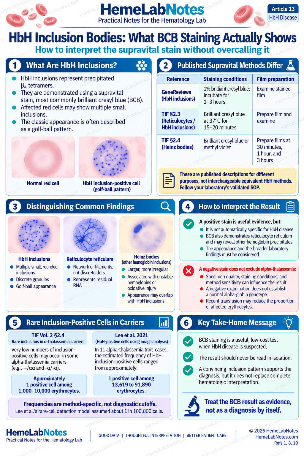
HbH inclusion staining is one of the most useful practical investigations when HbH disease is suspected.
Unlike a routine Wright–Giemsa blood film, this examination uses a supravital stain, commonly brilliant cresyl blue (BCB).
Under appropriate staining conditions, unstable HbH can precipitate within erythrocytes, producing multiple small inclusions.
The characteristic appearance is often described as a golf-ball pattern.
The term refers to the numerous stained inclusions visible within an affected red cell.
What the Stain Demonstrates
The characteristic HbH inclusions represent precipitated β₄ tetramers.
GeneReviews describes HbH inclusion-positive cells in approximately 5–80% of erythrocytes, following its specified BCB procedure. [1]
This is a broad range, and that matters.
The proportion of positive cells varies between cases and may be influenced by the method used.
It should not be converted into a universal positivity threshold or an independent measure of disease severity.
A convincing inclusion pattern is useful evidence, but the laboratory still needs to understand what its own method can reliably demonstrate.
Why the Incubation Method Matters
One detail worth examining is the variation between published BCB methods.
GeneReviews describes incubation with 1% brilliant cresyl blue for one to three hours when demonstrating HbH inclusions. [1]
The Thalassaemia International Federation (TIF) laboratory protocols describe another published procedure.
In Section 2.3, Reticulocyte and HbH Inclusion Body Detection, the BCB staining method includes incubation at 37°C for 15–20 minutes, followed by film preparation and microscopic examination. [8]
The same reference describes a different examination schedule in Section 2.4, Heinz Inclusion Bodies, with films prepared after 30 minutes, one hour, and three hours.
These protocols address related but not identical investigations.
Their incubation times should not be interpreted as interchangeable methods with established equivalent sensitivity for HbH detection.
The practical point is that staining conditions, examination procedures, and reporting criteria must be appropriate for the intended test.
Published protocols provide useful technical background, but they do not replace the laboratory’s validated SOP.
What I Look For Under the Microscope
I am not simply looking for one isolated dark granule.
The characteristic HbH pattern consists of multiple fine, rounded inclusions distributed within affected red cells.
But BCB is not exclusive to HbH.
It also demonstrates residual RNA in reticulocytes and can reveal precipitated hemoglobin in other settings.
Reticulocyte reticulum has a more filamentous or network-like appearance, whereas HbH inclusions generally appear as multiple discrete granules.
Heinz bodies associated with unstable hemoglobins or oxidative injury may have different appearances, although the features can overlap.
Strictly speaking, HbH precipitates themselves are a type of hemoglobin inclusion and can be described as Heinz-body-like. Morphology alone cannot establish the chemical identity of the precipitated hemoglobin.
A 2015 case report by Sridevi and colleagues demonstrated how HbH inclusions were mistaken for reticulocyte reticulum, resulting in a spuriously elevated manual reticulocyte count. The distinction became clear only after the preparation was reviewed in conjunction with the hemoglobin analysis. [9]
That example highlights something important about supravital staining.
Recognizing an intracellular structure is only the first step.
We still need to decide what the structure represents.
When reviewing the preparation, I would consider whether the inclusions are numerous and discrete, whether the appearance is consistent across affected cells, and whether the findings agree with the CBC, reticulocyte result, and other laboratory evidence.
A routine Wright–Giemsa film cannot replace an appropriate supravital examination.
Equally, the supravital stain should not be interpreted without the rest of the laboratory findings.
Rare Inclusion-Positive Cells Can Occur in Alpha-Thalassemia Trait
An inclusion-positive result is important, but it is not automatically equivalent to a diagnosis of HbH disease.
One reason is that rare inclusion-positive erythrocytes can occur in some alpha-thalassemia carrier states.
The TIF laboratory protocols discuss very low numbers of inclusion-positive cells in certain alpha-thalassemia carriers, including --/αα and -α/-α genotypes.
In the section dealing with Heinz inclusion bodies, the reported frequency is approximately one positive cell among 1,000–10,000 erythrocytes. [8]
A later image-analysis study by Lee and colleagues investigated rare HbH inclusion-positive cells in alpha-thalassemia trait.
In 11 trait cases, the estimated frequencies ranged from approximately one positive cell among 13,619 erythrocytes to one among 91,890 erythrocytes. [10]
The investigators also used an approximate frequency of one in 100,000 cells when developing their rare-cell detection model.
These observations are valuable, but they should not be combined into a single reference interval.
The TIF figures come from its discussion of hemoglobin precipitate inclusions, while the Lee study used a particular image-analysis method to estimate rare HbH inclusion-positive cells.
The techniques, cell-counting approaches, and definitions are not necessarily interchangeable.
What we can reasonably take from these findings is that rare inclusion-positive cells may occur in carrier states, whereas established HbH disease generally produces a much more conspicuous inclusion pattern.
Even then, the number of positive cells must be considered alongside the CBC, morphology, hemolysis findings, and hemoglobin analysis.
A rare positive cell is not the same as a convincing HbH disease phenotype.
Nor should the proportion of positive cells be used as a stand-alone severity score.
What If the Inclusion Test Is Negative?
The opposite situation deserves attention too.
Suppose the CBC shows marked microcytosis with more substantial anemia and reticulocytosis. The blood film contains anisopoikilocytosis, and the overall pattern raises concern about a clinically significant alpha-globin disorder.
But the HbH inclusion test is negative.
Should the investigation stop?
I would be cautious.
The sensitivity of an inclusion examination depends on the specimen, staining conditions, preparation quality, examination technique, and underlying phenotype.
A negative finding in one preparation does not establish a normal alpha-globin genotype.
The TIF laboratory protocols specifically caution that the absence of inclusions does not exclude alpha-thalassemia. [8]
This broader statement should not be treated as a precise measure of diagnostic sensitivity for every HbH disease subtype or staining method.
Recent transfusion may also complicate interpretation because donor erythrocytes can dilute the patient’s abnormal red-cell population.
If suspicion remains convincing, I would review the specimen and method, consider whether repeat examination is appropriate, and assess the hemoglobin analysis and molecular findings.
The decision should depend on the strength of the wider evidence.
A negative stain is a laboratory result that needs interpretation, not a substitute for interpretation.
What HPLC and Capillary Electrophoresis Add
Hemoglobin analysis provides another important part of the investigation.
HbH disease may produce an identifiable HbH fraction, and suitable HPLC or capillary electrophoresis methods can demonstrate abnormal hemoglobin patterns.
However, the analytical platform matters.
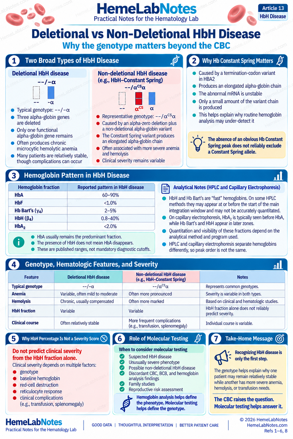
GeneReviews describes the following hemoglobin pattern in HbH disease. [1]
| Hemoglobin fraction | Reported pattern |
|---|---|
| HbA | 60–90% |
| HbF | <1.0% |
| Hb Bart’s | 2–5% |
| HbH (β₄) | 0.8–40% |
| HbA₂ | <2.0% |
These are published ranges, not mandatory diagnostic thresholds. The fractions vary between individuals and may be influenced by age, genotype, and analytical method. The stated ranges should not be interpreted as values expected to occur together in every sample.
One point worth remembering is that HbA usually remains the predominant fraction in HbH disease.
The presence of HbH does not mean HbA disappears.
Residual alpha-globin production still permits HbA formation, although the quantity is reduced.
This is an important distinction from Hb Bart’s hydrops fetalis syndrome, where the absence of functional alpha-globin production has very different consequences.
Why the Analytical Method Matters
On some HPLC systems, HbH appears as an early-eluting fraction.
The TIF laboratory protocols describe an HPLC procedure in which HbH and Hb Bart’s can be detected on the chromatogram but cannot be directly quantified because they elute before the start of integration. [8, Chapter 3, Section 3.1.1]
This is a method-specific limitation, not a property shared by every HPLC platform.
The same TIF reference notes that capillary electrophoresis and certain HPLC systems can detect and quantify HbH and Hb Bart’s when appropriate analytical methods and software capabilities are available. [8, Chapter 1, Section 1.1.2]
The laboratory should therefore know whether its particular method has been validated to identify HbH, quantify it, or both.
HbH is also unstable, making specimen freshness and handling important considerations.
If the CBC and inclusion findings strongly suggest HbH disease but the chromatogram is inconclusive, I would review the raw pattern, platform limitations, and specimen condition before dismissing the possibility.
An apparently unremarkable numerical report is not always equivalent to an unremarkable chromatogram.
HbH Is Not the Same as Hb Bart’s
The terminology is particularly important in newborn investigations.
HbH = β₄
Hb Bart’s = γ₄
Hb Bart’s is relevant during fetal and neonatal life because gamma-chain production predominates before the postnatal transition toward beta-chain synthesis.
Detecting Hb Bart’s in a newborn may provide evidence of alpha-globin deficiency.
However, detecting some Hb Bart’s is not equivalent to diagnosing Hb Bart’s hydrops fetalis syndrome.
The amount detected, the patient’s age, the complete hemoglobin pattern, and the underlying genotype all matter.
In adult HbH disease, the characteristic abnormal tetramer is HbH.
The two names should not be used interchangeably.
Deletional and Non-Deletional HbH Disease
Recognizing HbH disease is only part of the investigation.
The underlying alpha-globin defect helps explain why patients with apparently similar laboratory findings may have different clinical courses.
Deletional HbH Disease
The classic deletional genotype is:
--/-α
One chromosome has lost both alpha-globin genes, while the other has lost one.
Only one functional alpha-globin gene remains.
This produces reduced alpha-chain synthesis and allows excess beta chains to form HbH.
Deletional HbH disease often produces a relatively stable microcytic hemolytic anemia. Many patients do not require regular transfusions, although clinical complications can still occur. [3]
Some patients are diagnosed incidentally, while others present with anemia, splenomegaly, jaundice, or episodes of hemolytic worsening.
Infections and oxidative stress may precipitate acute deterioration.
Chronic hemolysis can contribute to gallstone formation. Iron overload may also develop even without regular transfusion because altered iron regulation and increased intestinal iron absorption can occur. [1,3]
This is another reason why microcytosis alone should not trigger iron supplementation.
Iron deficiency needs to be demonstrated.
A clinically mild HbH phenotype is not the same as an uncomplicated carrier state.
Non-Deletional HbH Disease
Not every HbH disease results from three conventional gene deletions.
Some patients inherit an alpha-zero deletion together with a non-deletional alpha-globin variant.
An important example is HbH–Constant Spring disease.
A representative genotype is:
--/αᶜˢα
Hb Constant Spring results from a termination-codon variant in the HBA2 gene.
The normal stop signal is altered, allowing translation to continue beyond the usual end of the alpha-globin chain.
This produces an elongated alpha-globin variant.
However, the abnormal mRNA is unstable and undergoes accelerated degradation, resulting in very low production of the variant chain.
Instability of the elongated globin product may also contribute to the disease process.
When this variant occurs alongside an alpha-zero deletion, the resulting HbH–Constant Spring phenotype is often more severe than typical deletional HbH disease. [4]
Patients may experience greater anemia, more pronounced hemolysis, splenomegaly, and increased transfusion requirements.
But that is a general tendency, not an absolute rule.
Non-deletional variants differ, and even individuals with similar genotypes may have different clinical courses.
The important distinction is that identifying HbH disease does not automatically define its molecular subtype.
The CBC, blood film, inclusion stain, and hemoglobin analysis can support the diagnosis.
Molecular testing helps establish which alpha-globin defect is responsible.
Why Hb Constant Spring Can Be Missed
Hb Constant Spring deserves particular attention because the abnormal hemoglobin fraction may be present at a very low concentration.
Depending on the analytical platform and underlying genotype, it may be difficult to recognize on routine hemoglobin analysis.
Therefore, the absence of an obvious Hb Constant Spring peak does not reliably exclude an underlying Constant Spring allele.
When the clinical and laboratory phenotype suggests a more severe alpha-globin disorder than the measured fractions explain, the limitations of the analytical method should be reviewed.
Molecular investigation may be necessary to resolve the discrepancy.
Do Not Predict Severity From HbH Percentage Alone
It can be tempting to interpret a higher HbH fraction as evidence of more severe disease.
But the relationship is not that straightforward.
Consider two patients with detectable HbH.
One has a higher HbH percentage but relatively stable anemia.
The other has a lower HbH percentage with more substantial anemia and clinical complications.
Which patient has the more severe disease?
The HbH percentage alone cannot answer that question.
A Thai study by Pornprasert and colleagues compared 43 deletional HbH cases with 22 non-deletional HbH–Constant Spring cases. [6]
The 43 deletional cases comprised 39 patients with the --SEA/-α3.7 genotype and four with --SEA/-α4.2.
All 22 non-deletional cases had the --SEA/αᶜˢα genotype.
The HbH–Constant Spring group generally showed more pronounced anemic features, including lower RBC counts and hemoglobin concentrations. The relationships between HbH levels and hematologic parameters also differed between the two groups.
This was a specific comparison between deletional HbH and HbH–Constant Spring disease. The findings should not automatically be extended to every possible non-deletional genotype.
Nevertheless, the study illustrates why the measured HbH fraction cannot be treated as a universal severity scale.
The patient’s hemoglobin concentration, reticulocytes, hemolysis findings, clinical history, and molecular genotype remain important.
The HbH fraction tells us something about the hemoglobin pattern.
It does not tell us everything about the patient.
Three Bench Examples
The following examples are illustrative teaching cases, not actual patient data.
Example A — A Pattern That Raises Suspicion of HbH Disease
| Parameter | Result |
|---|---|
| Hb | 9.8 g/dL |
| RBC | 5.25 × 10¹²/L |
| MCV | 62 fL |
| MCH | 18.7 pg |
| RDW | Increased |
| Reticulocytes | 4.2% |
The peripheral blood film shows marked microcytosis, hypochromia, anisopoikilocytosis, target cells, and polychromasia.
Iron studies do not support iron deficiency.
Further investigation identifies HbH inclusion bodies on supravital staining, and hemoglobin analysis demonstrates an HbH-containing fraction.
This is more than an isolated low MCV.
The combination of anemia, microcytosis, increased reticulocyte production, morphology, and HbH evidence strongly supports further investigation for HbH disease.
Molecular testing can help define whether the underlying disorder is deletional or non-deletional.
The important lesson is that the CBC raised the question, but the additional laboratory findings made the interpretation much stronger.
Example B — HbH Disease With Superimposed Iron Deficiency
A patient has previously confirmed HbH disease.
Earlier CBCs showed a stable hemoglobin around 10.7 g/dL, with persistent microcytosis and a longstanding thalassemia-type blood-film pattern.
The current CBC is different.
| Parameter | Current result |
|---|---|
| Hb | 8.7 g/dL |
| RBC | 4.70 × 10¹²/L |
| MCV | 59 fL |
| MCH | 18.5 pg |
| RDW | Increased from baseline |
Iron studies show reduced ferritin and transferrin saturation.
The patient already has an inherited red-cell disorder, but the current deterioration still requires explanation.
The evidence supports superimposed iron deficiency.
This is not a situation in which I should attribute every abnormal result to HbH disease.
Nor should I assume that a patient with HbH disease cannot become iron deficient simply because iron overload is a recognized complication of the disorder.
The previous CBCs help establish the chronic phenotype.
The current iron studies help explain what has changed.
A known diagnosis should not prevent us from recognizing a new problem.
Example C — More Severe Anemia With a Non-Deletional HbH Pattern
| Parameter | Result |
|---|---|
| Hb | 7.6 g/dL |
| RBC | 4.10 × 10¹²/L |
| MCV | 58 fL |
| MCH | 18.5 pg |
| RDW | Increased |
| Reticulocytes | Increased |
The peripheral blood film shows pronounced microcytosis, hypochromia, anisopoikilocytosis, and polychromasia.
HbH inclusions are identified, and hemoglobin analysis shows an HbH-containing fraction.
Molecular testing identifies an alpha-zero deletion together with an Hb Constant Spring allele.
The laboratory findings support HbH–Constant Spring disease.
The more substantial anemia is consistent with the tendency toward greater severity in some non-deletional HbH disorders.
However, the clinical course still cannot be predicted from the HbH percentage alone.
The molecular finding provides an explanation for the phenotype and information that routine hematology testing could not establish independently.
When Molecular Testing Becomes Important
Molecular investigation becomes particularly valuable when the clinical or laboratory question requires a definitive alpha-globin genotype.
This may include suspected HbH disease, unexpectedly severe anemia, conflicting inclusion and hemoglobin analysis findings, possible non-deletional variants, or family and reproductive investigations.
Alpha-thalassemia is genetically heterogeneous.
Some variants involve common deletions. Others involve less common copy-number changes, sequence variants, or regulatory abnormalities.
Available molecular approaches include gap-PCR, MLPA, targeted mutation assays, Sanger sequencing, and broader sequencing methods. [7]
These methods do not necessarily detect the same types of abnormalities.
A panel designed for common deletions may miss an uncommon non-deletional variant.
Sequence analysis may not identify every deletion or copy-number change.
Therefore, a report stating “molecular testing negative” is only meaningful when we know what the assay was designed to detect.
The laboratory should understand its test coverage, limitations, and whether additional investigation is required.
Molecular characterization is also important beyond the immediate diagnosis.
The exact parental alpha-globin genotypes influence reproductive risk, including the possibility of HbH disease or Hb Bart’s hydrops fetalis syndrome in offspring.
Those risks cannot be determined reliably from the MCV or HbH percentage.
Hemoglobin analysis helps define the hemoglobin phenotype. Molecular testing helps identify the underlying alpha-globin genotype.
A Practical Approach at the Bench
When the CBC raises concern about HbH disease, I would approach the findings in five stages.
This is an interpretation framework rather than a substitute for a laboratory diagnostic algorithm or SOP.
1. Recognize the Pattern
Start with the CBC.
Look at the hemoglobin, RBC count, MCV, MCH, RDW, and available analyser information.
Then review previous results.
Is the microcytosis longstanding? Has the hemoglobin changed? Does the current pattern resemble the patient’s usual baseline?
A change from baseline may be just as important as an abnormal result.
2. Look for Additional Processes
Review iron status and evidence of increased red-cell turnover.
Does iron deficiency explain part of the anemia?
Are the reticulocytes increased? Do bilirubin, LDH, haptoglobin, and other available findings support hemolysis?
The aim is to avoid assuming that every abnormality has the same cause.
3. Review the Morphology
Examine the peripheral blood film for microcytosis, hypochromia, target cells, anisopoikilocytosis, and polychromasia.
If HbH inclusion staining is indicated, follow the laboratory’s validated supravital staining procedure.
Distinguish reticulocyte reticulum from hemoglobin inclusions and consider other possible sources of intracellular staining.
A convincing pattern is useful, but the result must agree with the wider laboratory picture.
4. Evaluate Hemoglobin Analysis
Review the full hemoglobin pattern, not only the numerical fractions.
Is HbH identified?
Could an early-eluting fraction have been excluded from the usual integration?
Is the method validated to detect or quantify the suspected abnormal hemoglobin?
Could specimen instability or a low-abundance variant affect the result?
The analytical method matters as much as the reported numbers.
5. Resolve the Unanswered Questions
If the findings remain suspicious or discordant, consider what additional testing would clarify the diagnosis.
Molecular investigation may be appropriate to establish the alpha-globin genotype, especially when non-deletional disease, clinical implications, or family counseling are important.
Not every patient requires every investigation.
The purpose is to ensure that the next test answers a question the current evidence cannot settle.
Common Interpretation Traps
Trap 1 — A Very Low MCV Must Mean HbH Disease
Marked microcytosis is not specific.
Iron deficiency and other thalassemia syndromes can produce similarly low indices.
The complete CBC, reticulocytes, morphology, and relevant investigations determine whether HbH disease is a reasonable possibility.
Trap 2 — A High RBC Count Means It Is Only Thalassemia Trait
A relatively preserved RBC count supports a thalassemia-type pattern but does not establish clinical severity.
HbH disease can still be present.
The hemoglobin concentration and evidence of hemolysis remain important.
Trap 3 — Any HbH Inclusion-Positive Cell Proves HbH Disease
Rare inclusion-positive cells can occur in some alpha-thalassemia carriers.
The frequency and appearance of the inclusions, analytical method, and accompanying laboratory pattern matter.
A convincing HbH inclusion pattern is meaningful evidence, but it does not independently establish the genotype.
Trap 4 — Treating the BCB Result as Definitive
A positive supravital stain is not automatically specific for HbH.
BCB also demonstrates reticulocyte reticulum and may reveal other hemoglobin precipitates. The appearance of the inclusions and the accompanying laboratory findings must be considered.
Conversely, a negative examination does not establish a normal alpha-globin genotype.
Specimen quality, staining conditions, method sensitivity, and the underlying phenotype can influence the result.
A stain cannot provide more certainty than the method supports.
Trap 5 — A Higher HbH Percentage Means More Severe Disease
HbH percentage is not a stand-alone severity score.
Genotype, hemoglobin concentration, red-cell destruction, and clinical manifestations all contribute to the overall phenotype.
Trap 6 — Microcytosis Means the Patient Needs Iron
HbH disease produces microcytosis without necessarily involving iron deficiency.
Iron overload may occur, but superimposed iron deficiency is also possible.
Iron treatment should be based on demonstrated deficiency, not MCV alone.
Trap 7 — A Normal-Looking Hemoglobin Analysis Rules Out an Alpha-Globin Disorder
Routine hemoglobin analysis does not detect every alpha-globin abnormality.
HbH may be affected by pre-analytical and analytical limitations, while variants such as Hb Constant Spring may be present at low concentrations.
When the laboratory phenotype remains convincing, the discrepancy deserves investigation.
What the Laboratory Can Establish
The laboratory can identify a pattern suggesting HbH disease.
It can demonstrate marked microcytosis, hypochromia, and evidence of increased red-cell turnover.
The peripheral blood film may provide supporting morphology, while supravital staining and hemoglobin analysis can provide more specific evidence of HbH.
These findings can make a strong case for further investigation.
But an abnormal CBC index does not establish the molecular diagnosis.
An inclusion stain does not identify the precise genotype.
And an HbH percentage does not independently predict clinical severity.
Knowing these limitations is part of sound laboratory practice.
The goal is not to claim more than the evidence supports.
It is to recognize what the results tell us, what remains uncertain, and which investigation would meaningfully answer the next question.
The Takeaway
HbH disease can begin with a familiar laboratory picture: low MCV, low MCH, and a thalassemia-type RBC pattern.
But when the anemia is more pronounced, the reticulocyte count is increased, or the blood film suggests greater red-cell disturbance, the pattern deserves a second look.
HbH inclusion staining and hemoglobin analysis can provide important evidence. Molecular testing can then help explain the underlying alpha-globin defect and why disease severity differs between patients.
The aim is not to diagnose HbH disease from every markedly microcytic CBC.
It is to recognize when the findings tell us that there is more to investigate.
When microcytosis comes with evidence of hemolysis, do not stop at “thalassemia trait.” Read the rest of the pattern.
References
-
Tamary H, Greenberg-Kushnir N, Dgany O. Alpha-Thalassemia. In: Adam MP, Bick S, Mirzaa GM, et al., eds. GeneReviews® [Internet]. Seattle (WA): University of Washington, Seattle; 1993–2026. Updated April 23, 2026.
-
Makariou C, Papasavva T, Kleanthous M. Epidemiology, pathophysiology and diagnosis of α-thalassaemia. In: Amid A, Lal A, Coates TD, Fucharoen S, eds. Guidelines for the Management of α-Thalassaemia. Thalassaemia International Federation; 2023. Chapter 1.
-
Lal A. Clinical presentation and management of deletional haemoglobin H disease. In: Amid A, Lal A, Coates TD, Fucharoen S, eds. Guidelines for the Management of α-Thalassaemia. Thalassaemia International Federation; 2023. Chapter 2.
-
Songdej D, Teawtrakul N. Clinical presentation and management of non-deletional HbH disease. In: Amid A, Lal A, Coates TD, Fucharoen S, eds. Guidelines for the Management of α-Thalassaemia. Thalassaemia International Federation; 2023. Chapter 3.
-
Harteveld CL, Higgs DR. α-Thalassaemia. Orphanet J Rare Dis. 2010;5:13.
-
Pornprasert S, Salaeh N, Tookjai M, Punyamung M, Pongpunyayuen P, Treesuwan K. Hematological analysis in Thai samples with deletional and nondeletional HbH diseases. Lab Med. 2018;49(2):154–159.
-
Vijian D, Wan Ab Rahman WS, Ponnuraj KT, Zulkafli Z, Mohd Noor NH. Molecular detection of alpha thalassemia: a review of prevalent techniques. Medeniyet Medical Journal. 2021;36(3):257–269.
-
Old J, Harteveld CL, Traeger-Synodinos J, Petrou M, Angastiniotis M, Galanello R. Prevention of Thalassaemias and Other Haemoglobin Disorders: Volume 2: Laboratory Protocols. 2nd ed. Thalassaemia International Federation; 2012.
Chapter 1 — Best Practice Recommendations
https://www.ncbi.nlm.nih.gov/books/NBK190580/
Chapter 2 — Haematological Methods
https://www.ncbi.nlm.nih.gov/books/NBK190583/
Chapter 3 — Haemoglobin Pattern Analysis
-
Sridevi HB, Hegde A, Balanthimogru P, Khadilkar UN, Chakraborti S. Reticulum vs inclusions: a learning experience in haemoglobin H disease. J Clin Diagn Res. 2015;9(10):ED17–ED19.
-
Lee SY, Chen CME, Lim EYP, Shen L, Sathe A, Singh A, et al. Image analysis using machine learning for automated detection of hemoglobin H inclusions in blood smears: a method for morphologic detection of rare cells. J Pathol Inform. 2021;12:18.
Further Reading
Bain BJ, Bates I, Laffan MA. Dacie and Lewis Practical Haematology. 12th ed. Elsevier; 2017.
Keohane EM, Preston MM, Mirza KM, Walenga JM, eds. Rodak’s Hematology: Clinical Principles and Applications. 7th ed. Elsevier; 2024.
HemeLabNotes provides practical educational material for laboratory professionals. Interpretation and testing procedures should follow the laboratory’s validated methods, applicable standards, and clinical context.